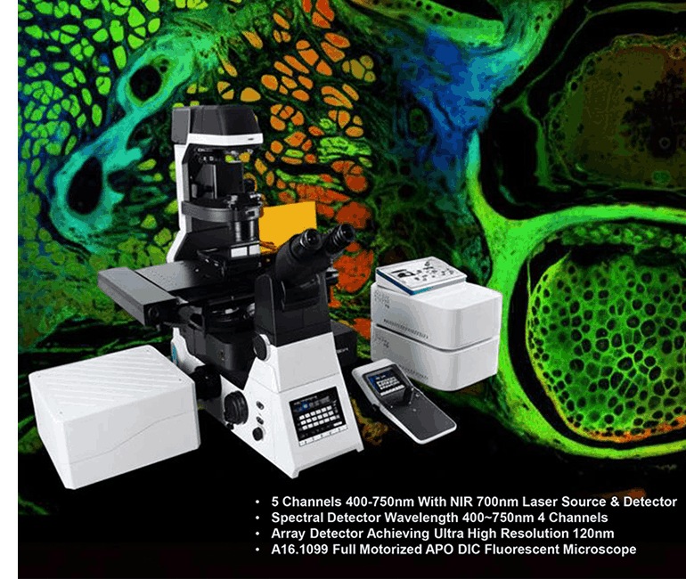
Clear images at high magnification now count as a basic need in both labs and factories. Older widefield systems run into trouble with thick samples. Light from planes above and below the focus point mixes together and hides fine detail. The confocal laser scanning microscope fixes this issue. It places a small pinhole in the light path so that only the in-focus plane reaches the detector. The OPTOEDU Maxcope A64 series takes the same idea and adds features that push 3D work further.
What is CLSM Microscopy and How Does It Redefine Clarity?
Switching from widefield to clsm microscopy brings a clear gain in sharpness. A laser beam lands on one thin layer at a time. The beam moves across the sample in small steps. Each step records only that single plane. The result shows structures that older tools miss.
Principle Behind Optical Sectioning and 3D Reconstruction
Optical sectioning forms the main working principle of the confocal laser scanning microscope. Standard microscopes light up the whole sample at once. This system uses a pinhole that sits at the image plane. Light from any other depth hits the pinhole edge and stays out. Only the chosen layer forms a clean, high-contrast picture. The Maxcope 2D/3D software then stacks these pictures taken at different depths. The stack builds a full 3D model. No cutting of the sample is required.
Why Multi-Mode Observation Matters for CLSM Microscopy
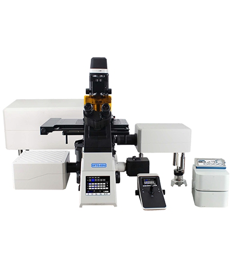
Professional work often needs several contrast methods on the same sample. The A64.1020 NIR model offers brightfield, phase contrast, polarizing, fluorescence, DIC, and Hoffman modes. Each mode brings out different sample features. Brightfield shows overall shape. Phase contrast makes transparent cells visible. DIC adds surface relief. Fluorescence tags specific molecules. The confocal laser scanning microscope can move between these modes on one stage position. Users therefore study cell shape, chemistry, and refractive index without moving the specimen.
Why Should You Invest in Confocal Laser Scanning Microscopy ( CLSM )for Your Lab?
Labs that need repeatable results and steady output look for systems that combine strong optics with solid automation. The OPTOEDU A64 series meets both needs. It gives fine detail and runs long imaging jobs with little operator input.
Breaking Depth Barriers with NIR Laser Technology
Visible light scatters fast inside thick tissue. The A64.1020 model uses near-infrared lasers instead. NIR light travels farther and causes less cell stress. In confocal laser scanning microscopy ( CLSM )this means clearer pictures from deeper layers. Researchers can watch live samples for longer periods without strong light damage.
Efficiency of Full Auto Motorized Systems in Confocal Laser Scanning Microscopy
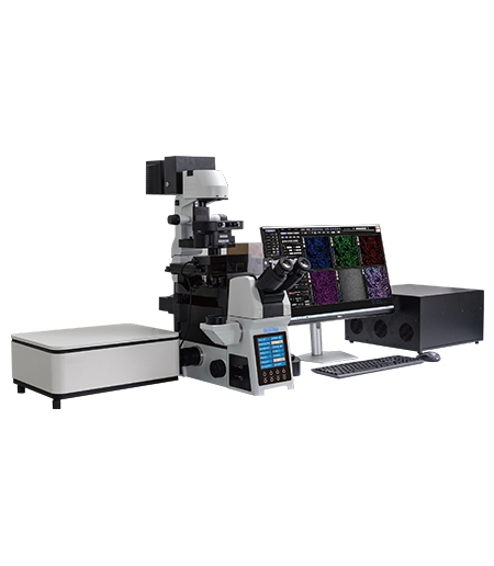
Motorized parts cut down on small positioning mistakes. The A64 series includes:
- XYZ stages that move the sample in exact steps for tiled scans.
- A motorized nosepiece that changes objectives on command.
- Motorized condensers and filter wheels that set the right light path for each mode.
With these parts the confocal laser scanning microscope can collect large image sets or Z-stacks on its own.
Superior Image Quality via APO Apochromatic Objectives
Lens quality sets the final limit on detail. The A64 series uses APO objectives. These lenses correct color and shape errors over a wide wavelength range. The laser spot stays tight and collected light stays in focus. Edge sharpness and contrast stay high even at 1000× magnification.
How a Confocal Laser Scanning Microscope Transforms Research and Industry
Many fields now depend on the level of detail that only clsm microscopy supplies.
From Biological Discovery to Live Cell Imaging
Pathology labs use the system to scan cervical cell slides. The motorized stage moves across the whole slide. The software stitches the fields into one large digital image. Doctors can zoom from the full tissue view down to single cells without losing focus. This helps spot early cancer signs more reliably.
How the Confocal Laser Scanning Microscope Excels in Industrial 3D Measurement
Factories also gain from 3D data. Typical tasks include:
- Checking wafer surfaces for small defects.
- Mapping chip circuit layers for failure analysis.
- Measuring resistor volumes in three dimensions.
The confocal laser scanning microscope does these jobs without touching or cutting the parts.
Seamless Large-Area Stitching with Advanced Maxcope Software
Large samples need many fields of view. The Maxcope software lines up overlapping images and blends them. Motor control keeps each new field in the right place. The final stitched file shows both the whole layout and the smallest details on one screen.
Conclusion
The confocal laser scanning microscope has become a standard tool for research and quality work. The OPTOEDU Maxcope A64 series adds NIR lasers, full motor control, and APO optics. Together these features let users see inside thick samples in three dimensions. Labs that choose a system with clsm microscopy and multiple contrast modes stay ready for new projects and tighter specs.
Contact us to learn more about how our professional microscope solutions can support your next project.
FAQ
Q: What is a confocal laser scanning microscope used for in research?
A: It captures thin optical sections at different depths. Stacking these sections gives a 3D view of cells, proteins, or industrial parts without cutting the sample.
Q: How does clsm microscopy improve 3D imaging over widefield?
A: Widefield systems collect light from every layer at once. The confocal laser scanning microscope blocks light from out-of-focus layers. The remaining signal is sharp, so 3D models stay clear.
Q: Why choose an OPTOEDU confocal laser scanning microscopy ( CLSM )system for industrial inspection?
A: The A64 series combines APO lenses, motorized stages, and stitching tools. These parts work together for fast, repeatable 3D checks on wafers, chips, and other small parts.
Q: Can a confocal laser scanning microscope perform multi-modal observations like DIC or Hoffman?
A: Yes. The A64.1020 model supports brightfield, phase contrast, fluorescence, DIC, and Hoffman modes. Users can switch modes on the same stage position to collect different types of data.
Q: What are the advantages of NIR laser in clsm microscopy?
A: NIR light passes deeper into tissue or materials. It scatters less and reduces cell stress. This makes long, live-cell studies and thick-sample inspection more practical.