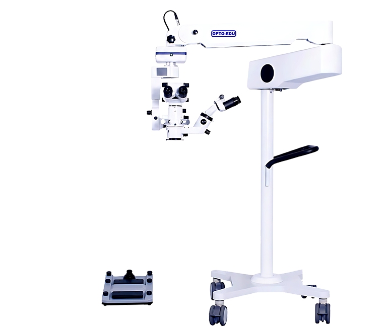
An ophthalmology operating microscope for cataract surgery should give the surgeon a stable red reflex, controlled illumination, smooth magnification changes, and hands-free positioning during delicate intraocular work. The red reflex is especially important because it helps reveal the lens, capsule, and surgical plane more clearly under coaxial light. OPTOEDU A41.1935 supports this workflow with motorized continuous zoom, dual-head observation, electromagnetic locks, a 14-function foot switch, and dual coaxial LED illumination for ophthalmic procedures.
The key to successful selection is how well the optical pathway, surgeon ergonomics, assistant view, and control system cooperate when the procedure requires focus. For cataract, retina, fundus, and vitreoretinal examination, a surgical microscope should provide the surgeon with a means of controlling the eye, the field, and the instrument.
Why an Ophthalmology Operating Microscope Depends on Red Reflex
Clearer Lens and Capsule Visualization
Red reflex is the bright retinal reflection seen through the pupil when coaxial illumination enters the eye and returns through the optical path. In cataract surgery, that reflection helps the surgeon judge contrast around the lens, capsule, and central working area. A weak or unstable reflex can make the field feel flat, especially when the lens opacity changes the way light returns to the observer.
A red reflex microscope is valuable because it supports visual confidence without forcing the surgeon to overcompensate with hand movement or excessive brightness. OPTOEDU A41.1935 is built for ophthalmology with red reflection and a macular protector.
Illumination That Supports Detail Without Distraction
Illumination quality matters as much as raw intensity. A41.1935 uses dual coaxial LED illumination with a listed brightness of 70,000 lux and aspheric multilayer-coated optics. In practical terms, coaxial light helps align the illumination with the viewing axis, which is important when surgeons need a clear reflection from deeper ocular structures.
The product also includes brightness control through the foot switch, allowing adjustment without leaving the operating position. That control is useful when the lens, cornea, or working fluid changes the optical condition.
Optical Control Beyond Basic Magnification
Motor Zoom for Continuous Field Adjustment
A cataract surgery microscope should move from overview to fine detail without breaking the surgeon’s rhythm. A41.1935 provides motorized continuous zoom from 4.5x to 27.3x with a parallel light optical system. Continuous zoom is helpful because the surgeon can refine the field gradually instead of jumping between fixed magnification steps.
During ophthalmic surgery, smooth magnification control can help maintain orientation while the surgeon follows small changes in the anterior or posterior segment field.
Dual-Head Observation for Surgeon and Assistant
The dual-head design supports observation for both the main surgeon and an assistant. OPTOEDU describes the main binocular tube and assistant tube as using independent optical systems with the same luminance and magnification. That matters in teaching, assisted surgery, and team-based procedures where both observers need a coherent view of the same surgical field.
The binocular tube also supports a 0-200 degree variable angle, which helps reduce long-session fatigue. In repeated ophthalmic work, ergonomics affects steadiness, posture, and focus.
How OPTOEDU A41.1935 Supports Ophthalmic Workflow
Hands-Free Control Through the Foot Switch
The A41.1935 foot switch brings key operations to the surgeon’s feet, including motorized zoom, motorized focus, X-Y movement, brightness adjustment, image capture, recording, picture browsing, freeze, light control, page movement, and filter switching. This is more than convenience. In ophthalmology, every unnecessary hand movement can interrupt the working posture or disturb a carefully maintained view.
For documentation-heavy workflows, image capture and recording control can also support case review. The product information also mentions a 4-inch touch screen and a foot pedal customization setting interface.
Electromagnetic Locks for Balanced Positioning
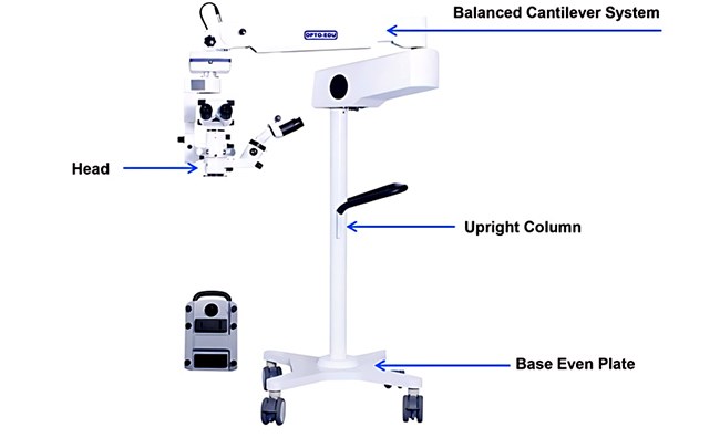
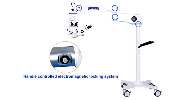
A surgical operating microscope must move freely when being positioned and then remain steady once the working area is found. A41.1935 uses three electromagnetic locks, and the handle button releases the lock system so the microscope lens can move to the working area. Releasing the button locks the joints again.
This free-floating and locked-position behavior supports fatigue-free surgery by balancing reach, movement, and stability. It is useful when the microscope must be repositioned while the field and assistant view remain coordinated.
Cataract, Fundus, and Retina-Oriented Use Scenarios
Cataract Surgery and Red Reflex Control
For cataract surgery, the microscope’s red reflex capability, coaxial illumination, and motorized zoom all work toward one goal: keeping the lens and intraocular structures readable. The surgeon needs contrast, depth impression, and a stable field while the procedure moves through different optical conditions. A41.1935 gives the operator tools to adjust zoom, focus, X-Y position, and brightness without changing hand position. The macular protector also belongs in this selection discussion because the retina must be protected during ophthalmic illumination.
Optional BIOM Kit for Fundus and Vitreoretinal Viewing
A41.1935 can also be paired with an optional BIOM kit. The non-contact wide-angle viewing system is described for fundus and vitreoretinal surgery, providing a panoramic high-resolution retinal view without corneal contact. This makes the system relevant beyond a narrow cataract-only discussion.
The usage scenario for ophthalmic surgery includes the vitreous, retina, and macular area. For hospitals, training centers, and specialist ophthalmic rooms, that means the microscope can be considered in a broader eye surgery workflow where anterior and posterior segment visibility both matter.
Choosing the Right Surgical Operating Microscope for Ophthalmology
Match Optical Features to the Procedure
A cataract-focused facility should look closely at red reflex, coaxial illumination, magnification control, and surgeon posture. A retina-oriented workflow should also evaluate wide-angle viewing options and how easily the microscope can be repositioned. The OPTOEDU A41.1935 ophthalmology operating microscope brings those considerations together in a configuration built around motor zoom, red reflex, dual-head observation, and electromagnetic positioning.
When comparing systems, avoid reducing the decision to a single number such as lux, zoom range, or weight. A useful ophthalmic surgical microscope must combine optical clarity, light control, mechanical stability, and intuitive control.
Consider Assistant View, Mounting, and Daily Handling
Assistant observation matters when the microscope is used for teaching, collaboration, or more complex ophthalmic cases. A dual-head system with matched luminance and magnification helps the second observer follow the same field rather than working from a compromised view. That is especially valuable when the assistant needs to anticipate instrument movement or understand the next step.
Conclusion
An ophthalmology operating microscope becomes valuable when it helps the surgeon see more clearly, move more calmly, and control the field without unnecessary interruption. OPTOEDU A41.1935 addresses cataract and ophthalmic surgery needs through red reflex support, motorized continuous zoom, dual coaxial LED illumination, dual-head viewing, electromagnetic locks, and a multifunction foot switch. Its optional BIOM kit also extends the discussion toward fundus and vitreoretinal observation, where stable wide-angle viewing can matter just as much as magnification.
Explore OPTOEDU surgical microscope solutions to match your ophthalmic workflow with the right microscope configuration.
FAQ
Q: What is an ophthalmology operating microscope used for?
A: An ophthalmic operating microscope is used to ensure magnified, lit, and stable visualization during eye surgery. In cases related to cataract and retina surgeries, the instrument ensures visualization of delicate structures, working distance, focus, and magnification control.
Q: Why is red reflex important in cataract surgery?
A: The red reflex allows the surgeon to distinguish between structures using the pupil by reflecting light off the retina. In cataract surgery, the red reflex can enhance visualization of the eye structures, particularly as changes in the field occur as a result of opacity or fluid movement. A red reflex microscope supports clearer orientation during delicate intraocular work.
Q: What does motor zoom add to an ophthalmic surgical microscope?
A: Motor zoom allows the user to adjust magnification smoothly without manually changing the optical setting. On A41.1935, the listed continuous zoom range is 4.5x to 27.3x. This helps the surgeon move from a broader overview to finer detail while preserving posture, field orientation, and procedural rhythm.
Q: Can A41.1935 support fundus or retina-related observation?
A: Yes, A41.1935 can be used with an optional BIOM kit described as a non-contact wide-angle viewing system for fundus and vitreoretinal surgery. It is intended to provide a panoramic, high-resolution retinal view without corneal contact. The exact configuration should be matched to the planned ophthalmic workflow and accessories.