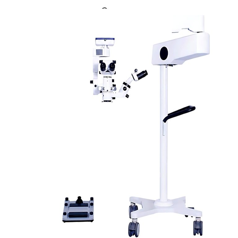
An ophthalmology operating microscope with motorized zoom should make it easier to move from a broad surgical view to fine detail without interrupting posture or hand position. OPTOEDU A41.1935 addresses that need with continuous 4.5x-27.3x motorized zoom, a parallel light optical system, dual-head observation, electromagnetic locks, and a foot switch that controls core microscope functions.
The useful question is not whether a microscope has a long specification list. It is whether its optical path, movement system, illumination, and controls cooperate when the surgeon needs a stable view of the eye. The OPTOEDU A41.1935 ophthalmology operating microscope is therefore best considered as a workflow tool for ophthalmic procedures rather than as a standalone magnification device.
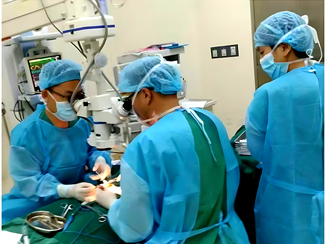
Why an Ophthalmology Operating Microscope Needs Motorized Zoom
Moving from Overview to Fine Detail
Eye surgery often requires more than one useful field of view. A wider view helps the surgeon maintain orientation, while higher magnification supports delicate work in a smaller area. A motorized zoom operating microscope allows those changes to happen continuously, so the image can be refined without a sudden jump between fixed steps.
A41.1935 provides a listed motorized continuous zoom range of 4.5x to 27.3x. That range gives the operator room to adapt the image to the procedure, the working depth, and the stage of observation while keeping the microscope aligned with the patient.
Keeping the Surgical Field Consistent
Manual changes to magnification can require a hand to leave its working position or can introduce a small pause while the field is re-established. Foot-controlled zoom keeps the adjustment separate from the instruments in the surgeon’s hands. The benefit is a smoother sequence of observation, positioning, and action.
The value of continuous control becomes clearer when the view must be adjusted several times during one procedure. Instead of treating magnification as a preset, the surgeon can use it as a live part of the visual workflow.
Optical Features That Shape Surgical Visibility
Parallel Light and Continuous Magnification
The A41.1935 optical design combines continuous motorized magnification with a parallel light system. For an ophthalmic surgical microscope, that combination matters because magnification is only useful when the field remains visually coherent as the operator changes scale.
A smooth zoom path can help preserve orientation when moving between the overall eye field and a more focused working area. It also reduces the need to repeatedly reach toward the optical head, which supports a steadier operating posture.
Red Reflection and Macular Protection
The microscope includes red reflection and a macular protector for ophthalmology. Red reflection can help the surgeon read contrast through the pupil during procedures where the lens and deeper ocular structures need to remain distinguishable. The macular protector belongs to the same optical conversation because light management and retinal awareness are connected to how the field is observed.
Illumination for Controlled Observation
A41.1935 uses dual coaxial LED illumination with brightness up to 70,000 lux. Coaxial illumination aligns the light more closely with the viewing path, while the aspheric multilayer-coated optics support light gathering and transmission.
The practical goal is controlled visibility, not brightness for its own sake. The foot switch allows brightness adjustment during the procedure, helping the operator respond when the visual condition of the field changes.
Control Without Breaking Surgical Posture
A Foot Switch for More Than Zoom
The surgical microscope foot switch on A41.1935 is designed to control motorized zoom, motorized focus, X-Y movement, brightness, image capture, recording, image browsing, freeze, light control, page movement, and filter switching. Keeping these actions at the feet can reduce the number of times the surgeon needs to leave the working posture.
This arrangement is especially useful when the surgeon needs to make a small visual correction while maintaining instrument awareness. It also gives the microscope a more consistent relationship with image documentation, because capture and recording controls are available within the same hands-free workflow.
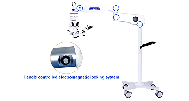
Electromagnetic Locks and Balanced Movement
The instrument uses three electromagnetic locks. Pressing the handle button releases the lock system so the microscope lens can be moved to the working area, and releasing the button locks the joints again. This free-floating and locked-position behavior supports quick positioning followed by stability.
For a motorized focus microscope, movement is only useful when it ends in a predictable position. The lock system helps separate repositioning from operation, allowing the microscope to move when needed and remain steady during observation.
Match the Microscope to the Procedure
Cataract and Anterior Segment Workflows
For cataract-related work, the selection focus should be on stable observation, controlled illumination, smooth magnification, and comfortable positioning. Red reflection, macular protection, motorized focus, and foot-controlled brightness can be considered together because each feature affects how the surgeon maintains visual orientation.
Fundus and Vitreoretinal Observation
A41.1935 can be paired with an optional BIOM kit for fundus and vitreoretinal surgery. The non-contact wide-angle system provides panoramic, high-resolution retinal viewing without corneal contact. That makes the instrument relevant as a fundus surgery microscope when the intended workflow extends beyond anterior segment observation.
The important selection question is whether the facility needs one microscope configuration to support both routine ophthalmic observation and posterior-segment viewing. The answer depends on the planned procedures and the accessory configuration, not on magnification alone.
How to Evaluate an Ophthalmology Operating Microscope
Look at the Whole Control Chain
When comparing an ophthalmology operating microscope, review the sequence from illumination to viewing, zoom, focus, movement, locking, and documentation. A strong specification in one area cannot compensate for a control layout that interrupts posture or an optical path that becomes difficult to manage when the view changes.
Consider the Assistant and Documentation Workflow
A dual-head system can support assistant observation and teaching, while image capture and recording can support review after the procedure. These features are more useful when they fit the room’s actual habits. The microscope should be evaluated alongside the people who position it, operate it, teach with it, and review the resulting images.
Why OPTOEDU A41.1935 Fits Motorized Ophthalmic Workflows
OPTOEDU A41.1935 brings continuous motorized zoom, dual coaxial LED illumination, red reflection, macular protection, electromagnetic positioning, dual-head observation, and foot-controlled operation into one ophthalmic platform. Its optional BIOM kit also gives specialist rooms a path toward non-contact fundus and vitreoretinal viewing.
The strongest use case is a facility that values controlled visual transitions and hands-free adjustment during ophthalmic work. Explore OPTOEDU surgical microscope solutions to compare the A41.1935 configuration with the demands of your operating workflow.
Conclusion
An ophthalmology operating microscope becomes more useful when magnification, focus, illumination, and positioning work as one system. OPTOEDU A41.1935 supports that approach with 4.5x-27.3x motorized continuous zoom, red reflection, macular protection, dual-head observation, three electromagnetic locks, dual coaxial LED illumination, and a multifunction foot switch.
Contact us to discuss how the OPTOEDU A41.1935 can support your ophthalmic surgical requirements.
FAQ
Q: What does motorized zoom add to an ophthalmology operating microscope?
A: Motorized zoom lets the surgeon move continuously between a broader overview and a more detailed field without manually changing the optical setting. On A41.1935, the listed range is 4.5x to 27.3x. Foot control helps preserve posture and keeps both hands available for the surgical task.
Q: Why is a foot switch important in ophthalmic surgery?
A: A foot switch keeps frequently used controls close to the surgeon’s operating position. A41.1935 uses it for zoom, focus, X-Y movement, brightness, image capture, recording, freeze, light control, and filter switching. This can reduce unnecessary hand movement during delicate visual work.
Q: Can A41.1935 be used for fundus and vitreoretinal procedures?
A: The microscope can be paired with an optional BIOM kit described for fundus and vitreoretinal surgery. The BIOM system provides non-contact wide-angle retinal viewing. The final configuration should be matched to the facility’s planned procedures and the accessories required for those procedures.
Q: How should a facility compare motorized ophthalmic microscopes?
A: Compare the complete workflow rather than one number. Review zoom range, focus, illumination, red reflection, positioning locks, assistant viewing, foot controls, image capture, and accessory compatibility. A microscope is a better fit when these elements support the surgeon’s posture and the room’s daily operating routine.