Microbiology laboratory solutions increasingly depend on imaging systems that can do more than provide a single microscopic view. For laboratories working with fluorescence imaging, live-cell observation, automated image acquisition, or multidimensional analysis, the right microscope should connect optical performance with stable positioning, time-based recording, and practical data management. OPTOEDU addresses these needs through motorized fluorescence microscopy, live-cell imaging, and confocal systems designed for different levels of biological research.
The most effective solution depends on what the laboratory needs to observe and how the resulting images will be used. A routine fluorescence workflow may benefit from automated positioning, while long-term live-cell work requires focus stability and controlled observation; more complex experiments may need confocal optical sectioning and multidimensional acquisition.
Microbiology Laboratory Solutions for Modern Imaging Workflows
A microscopy system becomes more valuable when its configuration reflects the experimental workflow rather than simply offering higher magnification. In microbiology laboratories, researchers may need to move between direct observation, fluorescence imaging, repeated positions, long-term monitoring, and digital analysis without rebuilding the imaging setup for every task.
OPTOEDU provides several current platforms that address different stages of this workflow. The A16.1095 focuses on motorized upright fluorescence imaging, the A16.1099 combines an inverted platform with functions for live-cell observation, the M30.4810 adds scheduled live-cell monitoring and data review, and the A64.1010 extends imaging into automated confocal and multidimensional acquisition.
Fluorescence Imaging With Motorized Control
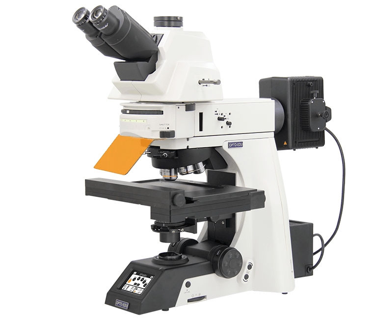
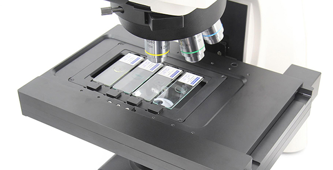
The OPTOEDU A16.1095 is an upright fluorescence microscope built around automation of routine microscope movements. Its epi-fluorescence system incorporates a noise-elimination design intended to produce bright, high-contrast images, while the full-auto X/Y/Z stage allows specimen movement to be controlled through the platform software and joystick.
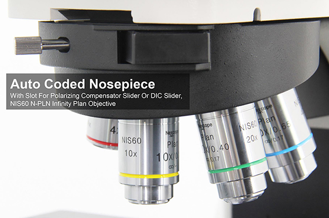
This type of motorization matters when a microbiology microscopy workflow requires repeated examination of different areas of a specimen. Instead of relying entirely on manual repositioning, the operator can control the stage systematically and change objectives through the coded nosepiece, helping integrate observation with digital image acquisition.
The A16.1095 also includes automated condenser movement and controls that allow focusing speeds to be set for different operating needs. These features do not replace the decisions of an experienced microscopist, but they reduce the number of repetitive mechanical adjustments involved in moving between fields, objectives, and imaging conditions.
Moving From Observation to Live-Cell Imaging
A static image provides information about one moment. When an experiment depends on changes occurring over hours or days, live-cell imaging requires a different approach because focus stability, environmental disturbance, and repeated acquisition become part of the imaging problem.
Maintaining Focus During Long-Term Observation
The OPTOEDU A16.1099 is an inverted fluorescence microscope designed specifically around demanding living-cell microscopic experiments. Its Adaptive Focus Shift system is intended to counter focus drift, while the live-cell culture system and high-brightness LED fluorescence source support multi-day time-lapse observation.
For microbiology laboratory solutions involving continuous biological observation, this changes the role of the microscope. Instead of repeatedly returning to the instrument to find the same region and restore focus, researchers can build an imaging workflow around more stable long-term acquisition.
The inverted configuration also fits workflows where specimens remain in culture vessels during observation. High-speed electric control coordinates objective changes, filters, the XY stage, and observation modules, while the system also supports DIC imaging for samples where enhanced contrast and structural detail are important.
Reducing Disturbance During Scheduled Imaging
The OPTOEDU M30.4810 Live Cell Imaging System approaches time-based experiments from another direction. Its software supports remote control, photo and video capture, time-lapse photography, cell counting, cell confluence analysis, scratch experiments, and email reminders, while motorized autofocus is included in the listed configuration.
Scheduled observation can be particularly useful when repeated manual access would interrupt an experiment. The M30.4810 is designed so that cell progress can be monitored remotely without repeatedly entering the clean area or opening the incubator, reducing disturbance to the growth environment and lowering the risk of sample contamination.
Its value continues after acquisition. Delayed-shooting experiments can be reviewed through data-analysis curves, recognized images, and time-lapse videos, so researchers can examine a sequence as an experimental record rather than treating each captured frame as an isolated image.
Automation Makes Microbiology Imaging More Repeatable
Automated microscopy for microbiology is most useful when an experiment requires many positions, repeated time points, or frequent changes in imaging conditions. Motorized stages and objective switching reduce repetitive manipulation and create a more structured relationship between specimen position and image acquisition.
That distinction becomes important when choosing between the A16.1095 and A16.1099. The upright A16.1095 is better aligned with automated fluorescence observation where the emphasis is on controlled stage movement and efficient acquisition, while the inverted A16.1099 adds features specifically designed around live-cell stability and extended time-lapse work.
For experiments in which continuous monitoring is the dominant requirement, the M30.4810 provides a more dedicated live-cell workflow. Its combination of scheduled observation, remote operation, autofocus, time-lapse recording, and software-based data review makes the imaging process less dependent on constant operator presence.
When Confocal Microscopy Adds More Information
Widefield fluorescence imaging is appropriate for many laboratory tasks, but complex research may require optical information from different depths, fluorescence channels, positions, and time points. This is where confocal microscopy becomes relevant as part of advanced microbiology laboratory solutions.
Combining Space, Wavelength, and Time
The OPTOEDU A64.1010 supports combined X, Y, Z, wavelength, and time scanning. Its acquisition modes include multi-channel fluorescence imaging, time-lapse scanning, multi-position imaging, Z-axis stacking, and panoramic stitching, and these modes can be combined according to experimental requirements.
This makes the system useful when a single two-dimensional fluorescence image does not contain enough information. A researcher can construct an acquisition around depth, multiple fluorescence signals, different specimen locations, or changes over time rather than repeatedly collecting unrelated images.
The system uses an integrated confocal pinhole design to filter non-focal-plane signals while improving fluorescence detection and image resolution. Its current configuration supports four-channel fluorescence detection, and the product catalogue also describes real-time synchronized four-channel imaging for observing multiple fluorescent labels in the same field.
Managing Larger and More Complex Image Sets
Advanced imaging also creates a data-management challenge. The A64.1010 uses the NomisProX-C software environment to integrate microscope hardware control with confocal imaging and analysis, reducing the separation between instrument operation and image processing.
The platform supports a 25 mm confocal field of view and scanning up to 8192 × 8192 pixels. Its catalogue also describes high-speed imaging of up to 60 fps under a specified 8 × 256-pixel scanning condition, making acquisition speed another consideration when experiments involve dynamic samples or repeated imaging.
Choosing the Right Microscope for a Microbiology Laboratory
Match the System to the Observation Method
The first selection question should be the type of observation required. If the workflow is centered on automated upright fluorescence imaging and systematic positioning, the A16.1095 provides motorized XYZ movement and fluorescence-oriented imaging functions without introducing a dedicated long-term cell culture workflow.
When experiments require extended observation of living cells, the A16.1099 is more closely aligned with that need because focus-drift compensation and a live-cell culture environment are part of its design. If unattended scheduled monitoring, remote access, and time-lapse data review are priorities, the M30.4810 provides a more specialized solution.
Consider the Complexity of Image Acquisition
Not every laboratory requires confocal microscopy. The A64.1010 becomes more relevant when research needs multi-channel fluorescence, Z-stacks, multiple positions, time-series acquisition, panoramic imaging, or combinations of these dimensions.
This approach helps laboratories avoid selecting equipment solely by technical sophistication. OPTOEDU microbiology imaging systems can instead be considered according to the actual progression of an experiment: how the sample is held, how long it must be observed, how frequently images are captured, whether fluorescence channels must be combined, and how much image analysis follows acquisition.
Conclusion
Modern microbiology laboratory solutions increasingly connect microscopy with automation, stable live-cell observation, time-lapse acquisition, fluorescence imaging, and multidimensional analysis. The value of these technologies lies not in adding automation for its own sake, but in helping researchers build an imaging workflow that matches the biological experiment and preserves useful information from observation through analysis.
OPTOEDU offers different paths for those requirements, from the motorized fluorescence workflow of the A16.1095 and the live-cell capabilities of the A16.1099 to scheduled monitoring with the M30.4810 and multidimensional confocal imaging with the A64.1010. Choosing among them should begin with the sample, observation duration, imaging mode, acquisition frequency, and level of analysis the laboratory actually requires.
Explore OPTOEDU microbiology laboratory solutions to connect fluorescence imaging, live-cell observation, automation, and advanced analysis within a more integrated laboratory workflow.
FAQ
Q: What should a microbiology laboratory consider when choosing a microscope?
A: The laboratory should first define its specimen format, required observation method, imaging duration, and data needs. Routine fluorescence imaging may benefit from motorized positioning, while living-cell experiments may require focus-drift control and time-lapse acquisition. Confocal microscopy becomes more relevant when the experiment requires Z-stacks, multiple fluorescence channels, multiple positions, or multidimensional analysis.
Q: What is the difference between the OPTOEDU A16.1095 and A16.1099?
A: The A16.1095 is an upright motorized fluorescence microscope with automated XYZ stage movement, objective switching, and fluorescence-oriented imaging functions. The A16.1099 is an inverted full-auto fluorescence system designed around living-cell experiments, adding Adaptive Focus Shift control, a live-cell culture system, and support for multi-day time-lapse observation.
Q: How does the M30.4810 support live-cell imaging workflows?
A: The M30.4810 supports scheduled observation, remote control, autofocus, photo and video capture, time-lapse imaging, cell counting, confluence analysis, and experimental data review. Researchers can examine analysis curves, recognized images, and delayed videos after acquisition, making it suitable for workflows where long-term monitoring and reduced manual intervention are important.
Q: When should a microbiology laboratory consider a confocal microscope?
A: A confocal system is worth considering when experiments require information beyond a conventional two-dimensional fluorescence image. The OPTOEDU A64.1010 supports multi-channel fluorescence, Z-axis stacking, time-lapse scanning, multi-position acquisition, panoramic stitching, and combined X, Y, Z, wavelength, and time imaging, allowing complex experiments to be organized within one acquisition environment.