A laser scanning confocal microscope creates sharper fluorescence images by using a pinhole to reject out-of-focus light, then scanning the sample layer by layer for optical sectioning and 3D reconstruction. For researchers working with living cells, tissue sections, organoids, coatings, or semiconductor surfaces, the value is not only higher magnification. It is cleaner signal, controllable depth, and the ability to connect multi-channel fluorescence with Z-stack imaging in a repeatable workflow.
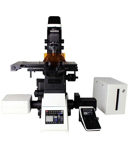
The OPTOEDU A64 confocal microscope family fits this need from different angles. A64.1020 emphasizes NIR observation, array detection, and 120 nm ultra-high resolution. A64.1010 focuses on automated 8K scanning and multi-dimensional acquisition, while A64.0960 provides a motorized scanning platform with 405, 488, 561, and 640 nm laser lines.
Why a Laser Scanning Confocal Microscope Sees More Than Widefield
The Pinhole That Creates Optical Sectioning
In widefield fluorescence microscopy, the entire illuminated thickness of the sample can contribute light to the image. That is fast and useful for simple specimens, but thick tissue, dense cell cultures, and uneven materials can quickly produce haze. A laser scanning confocal microscope narrows the imaging path so light from the focal plane is favored while unfocused fluorescence is filtered before detection.
The pinhole is the small optical gate that makes this possible. Excitation light and emitted sample light pass through a conjugate path, so the detector receives a cleaner slice of information instead of a blurred mixture from multiple depths. This is why pinhole optics matter for immunofluorescence localization, neuronal tissue sections, organoids, and surface layers where the position of a signal is as important as its brightness.
Cleaner Fluorescence Background for Complex Samples
Lower background changes how users interpret an image. A membrane marker, a nuclear stain, or a labeled cytoskeletal structure can be separated from surrounding fluorescence with better contrast. In materials work, the same principle helps distinguish surface features, transparent layer boundaries, or defects that ordinary fluorescence may blur.
OPTOEDU A64.1010 and A64.0960 both use motorized pinhole design, which keeps optical sectioning practical during repeated imaging tasks. Instead of treating confocal adjustment as a manual step, the system supports consistent transitions between sample types, magnifications, and fluorescence channels. That consistency matters when a lab compares multiple slides, time points, or sample regions.
Laser Lines and NIR Imaging in Real Fluorescence Workflows
Matching Wavelengths to Fluorophores
Confocal imaging depends on matching laser excitation to the fluorophore or stain. Common biological workflows often use 405 nm for DAPI or Hoechst nuclear staining, 488 nm for GFP or FITC, 561 nm for RFP or mCherry, and 640 nm for Cy5 or Alexa 647 style far-red labels. When those channels are separated clearly, multi-color imaging becomes easier to analyze without confusing one signal for another.
The OPTOEDU A64.0960 laser confocal scanning microscope is especially straightforward for this wavelength discussion because it includes 405 nm, 488 nm, 561 nm, and 640 nm lasers, with AOTF wavelength control across 400-750 nm. In a biological laboratory, this supports routine multi-channel fluorescence observation. In materials and biophotonics work, it also helps users choose excitation conditions for labeled structures or photoresponsive samples.
Where NIR Confocal Imaging Adds Value
NIR confocal microscope workflows are valuable when the sample benefits from longer-wavelength observation. Near-infrared light can help with deeper regions, lower scattering, and gentler observation when the sample and probe are suitable. For live-cell or tissue-oriented work, this can help reduce the pressure between signal strength and sample stress.
The OPTOEDU A64.1020 NIR laser confocal microscope combines NIR positioning with 5-channel imaging, 400-750 nm spectral detection, a 4-channel PMT system, and an array detector. Its 120 nm ultra-high resolution is relevant for fine intracellular or subcellular structures. The practical benefit is a platform that can move from routine fluorescence channels into more demanding low-signal or deeper-sample observation.
Z-Stack Imaging and 3D Reconstruction in Practice
From Optical Slices to a 3D Volume
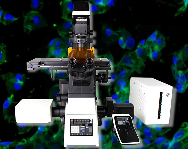
Z-stack imaging takes a series of optical sections at different focal depths and turns them into a depth-aware dataset. Instead of judging a thick sample from one plane, the user can follow structure through the sample and reconstruct spatial relationships. This is central to confocal applications in tissue sections, spheroids, organoids, plant samples, and developmental model organisms.
The workflow is also useful outside life science. Transparent coatings, layered films, micro-patterned surfaces, and semiconductor features may need depth-related information rather than a single top-down image. When combined with multi-site scanning or panoramic stitching, Z-stack imaging can connect local detail with a broader sample map.
Live Cells, Tissue Sections, and Organoids
Live-cell imaging needs speed and careful light exposure. A64.1020 uses high-speed resonant scanning to shorten illumination time while maintaining image quality, a combination that helps reduce photobleaching and phototoxicity during prolonged observation of sensitive samples. This is useful when researchers need time-lapse imaging of cell movement, protein transport, or structural change rather than one static snapshot.
For larger biological structures, OPTOEDU A64.1010 supports X, Y, Z, lambda, and T scanning combinations, with multi-channel fluorescence, time-lapse, multi-position acquisition, Z-axis stacking, and panoramic stitching. That combination matches how modern biology often works: locate the region, capture multiple signals, follow change over time, and build a 3D view when depth matters.
Choosing an OPTOEDU A64 Confocal Workflow
A64.1020 for NIR and High-Resolution Detection
A64.1020 is the strongest fit when the sample demands deep, low-signal, or high-resolution observation. Its array detector captures weak fluorescence signals, while the multi-channel detection structure supports flexible fluorescence experiments. The 5.7-inch touch screen, motorized nosepiece, motorized XYZ stage, motorized condenser, and motorized fluorescence turret also reduce manual interruptions during complex imaging sessions.
A64.1010 for Automated 8K Multi-Dimensional Imaging
The OPTOEDU A64.1010 laser confocal microscope suits laboratories that need high-throughput imaging with strong automation. Its 8K x 8K scan head, 4 laser sources, 4 PMT detectors, motorized pinhole, and controlled light-splitting modes support experiments that move between overview imaging and detailed fluorescence capture. For repeated sample comparison, automation is not a luxury; it helps keep the imaging process consistent.
Software integration also affects daily use. A64.1010 is built around a confocal software platform that connects microscope hardware control with confocal imaging analysis. That matters when the operator needs to switch objectives, adjust observation modes, acquire Z-stacks, manage time-lapse sequences, and visualize results without breaking concentration at every step.
A64.0960 for Motorized Confocal Scanning Applications
A64.0960 is a practical option when a lab needs a full motorized inverted fluorescence platform for scanning applications. Its scan modes include X-Y, X-Y-Z, and X-Y-Z-T, and its 16-bit image depth supports richer signal gradation in fluorescence work. The configuration also includes a computer, 4K monitor, digital camera, and professional 3D software for a complete confocal scanning station.
The application range is broad but still specific. In model animals and plants, wide field of view and layer scanning help reveal structures at different depths. In biomaterials research, the system can observe interactions between materials and living cells when paired with the appropriate living-cell environment monitoring module.
Application Scenarios Where Confocal Detail Matters
Life Science Imaging
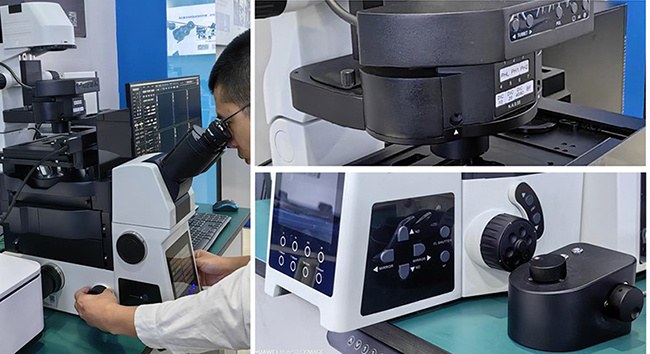
In cell biology, confocal microscopy helps show whether a fluorescent signal is inside the nucleus, along the membrane, near the cytoskeleton, or distributed across a tissue layer. That spatial clarity is difficult to obtain from a thick or bright sample using widefield alone. OPTOEDU A64 systems support these workflows through multi-channel fluorescence, optical sectioning, Z-stack acquisition, and motorized positioning.
Materials and Industrial Research
Confocal imaging also has a place in materials and industrial laboratories. Transparent film thickness, coating uniformity, surface roughness, and fine defect inspection often require depth-related imaging or cleaner optical contrast. A motorized laser scanning confocal microscope can help users return to the same region, compare layers, and document surface behavior with more structure than a simple 2D image.
Conclusion
A laser scanning confocal microscope becomes most valuable when pinhole optics, wavelength selection, scanning automation, and 3D reconstruction work together. Widefield imaging remains useful for fast observation, but confocal microscopy brings cleaner background, optical sectioning, multi-channel control, and Z-stack depth to samples that are too complex for a single focal plane. OPTOEDU A64.1020, A64.1010, and A64.0960 give laboratories different paths into that workflow, from NIR high-resolution imaging to automated 8K scanning and motorized 3D confocal applications.
Explore the OPTOEDU A64 confocal microscope solution that matches your live-cell, tissue, materials, or 3D imaging workflow.
FAQ
Q: What does a pinhole do in a laser scanning confocal microscope?
A: The pinhole blocks much of the out-of-focus light before it reaches the detector. This makes the image cleaner and allows the microscope to capture optical sections from selected depths. By stacking these sections, users can build a 3D view of thick biological samples, transparent layers, or structured materials.
Q: When is an NIR confocal microscope useful?
A: NIR confocal imaging is useful when longer-wavelength observation is suitable for the sample and fluorescent probe. It can support deeper imaging strategies, lower scattering conditions, and gentler observation of sensitive specimens. OPTOEDU A64.1020 is relevant for this workflow because it combines NIR positioning with array detection and high-resolution fluorescence capture.
Q: How is confocal microscopy different from widefield fluorescence microscopy?
A: Widefield fluorescence collects light from a larger illuminated volume, which is fast but can create haze in thick or bright samples. Confocal microscopy scans the sample and uses a pinhole to reject out-of-focus light. The result is cleaner optical sections, stronger depth control, and better support for Z-stack reconstruction.