Getting sharp, detailed images of thick biological samples remains a tough task. Standard widefield systems often pick up stray light from areas outside the focal plane, which creates unwanted blur. This problem has pushed imaging tools forward from the basic light scanning microscope toward the more precise laser confocal scanning microscope. Labs that need to see fine cellular structures must understand how these two systems differ in real performance.
OPTOEDU now offers equipment that lights the sample point by point. The design cuts background noise and improves the way optical sections are taken.
Redefining Clarity through the Laser Confocal Microscope
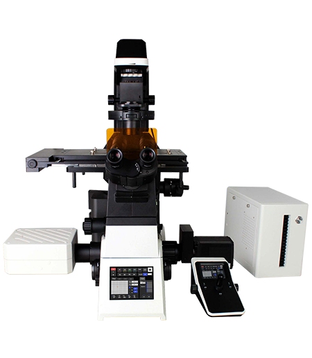
The key point is the way each device deals with the light before it gets to the sensor. The conventional method illuminates the entire sample at once, leading to much scattering. In the laser confocal microscope, a better optical path is formed, leaving just the focal plane in focus.
The Conjugate Pinhole Principle and Eliminating Blur
Every laser confocal scanning microscope contains a pinhole unit. The excitation light and the light coming back from the sample travel through the same small opening. Because the two paths stay conjugate, only the signal that starts at the focal plane reaches the detector. Light from other planes gets blocked. This raises both sensitivity and image sharpness.
Point-by-Point Imaging vs Widefield Illumination
Widefield systems record the full field in one shot. A laser confocal scanning microscope scans the sample point by point with a focused laser. The collected data is later rebuilt into a full picture. Because the laser hits only one small spot at a time, scattered light stays low even in dense tissue or thick model organisms.
Core Benefits of the Laser Confocal Scanning Microscope
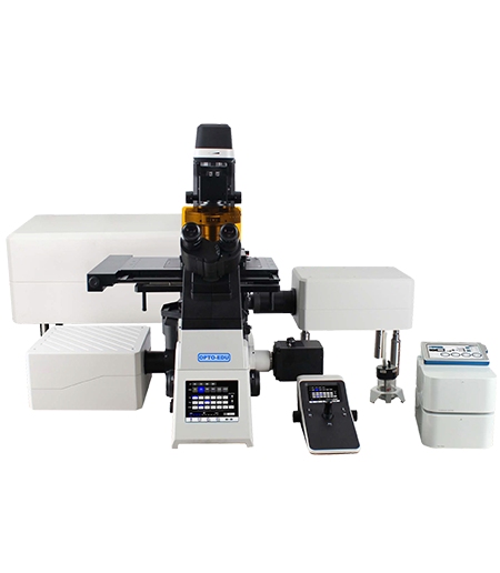
High-end work is judged by three things: resolution, imaging depth, and how well the sample stays healthy. OPTOEDU systems are built to meet all three needs at once.
Achieving 8K Resolution and Capturing Subtle Structures
The OPTOEDU A64.1020 uses a fast optical scanning galvanometer. It can record images at 8192 × 8192 pixels. At this 8K level, every small texture and cell detail stands out clearly. Researchers who study large structures or fast biological events now have a data set that a light scanning microscope cannot provide.
3D Reconstruction for Organoids and Thick Specimens
A laser confocal scanning microscope can scan layer by layer along the Z-axis. Software then builds a three-dimensional model from these optical sections. The method works well for organoids, cell clusters, and thick tissue slices. The A64.0960 has already produced clear 3D views of zebrafish embryos and Arabidopsis roots, showing spatial details that disappear in ordinary 2D widefield images.
Choosing Advanced Laser Systems over Standard Light Scanning
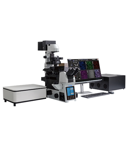
The choice between a light scanning microscope and a laser confocal system often depends on the exact biological question. Live-cell work and multi-protein studies place extra demands on speed, sensitivity, and sample safety.
Sensitivity and Speed in Live Cell Observation
Live-cell imaging needs both fast capture and low light damage. The OPTOEDU A64.1020 uses resonant scanning. This method runs nearly twenty times faster than earlier point-scanning designs. Because the laser spends less time on each spot, cells suffer less photobleaching and phototoxicity. GaAsP PMT detectors also collect more of the weak fluorescence signal that standard detectors often miss.
Simultaneous Multi-Channel Imaging and Colocalization
Watching several proteins at once requires good spectral control. The A64.1095 carries four integrated lasers at 405 nm, 488 nm, 561 nm, and 640 nm, plus separate detectors for each color. Four fluorescence channels can be recorded together, which supports accurate colocalization measurements. The software sets the best pinhole size for each objective’s numerical aperture, keeping the signal-to-noise ratio high in every channel.
Comparing Top Solutions: A64.1020 and A64.0960
Different labs face different imaging tasks. OPTOEDU supplies two main platforms so users can match the tool to the sample.
The NIR Advantage for Deep Tissue Imaging
Thick tissue often limits light penetration. The A64.1020 offers an optional 730 nm near-infrared laser and matching detector. Near-infrared light travels farther with less scatter, which helps when the target sits deep inside a tissue section or whole animal. Spectral unmixing tools then separate signals that have similar colors but different emission spectra, giving clearer qualitative results.
Scalability and Modular Application Expansion
A research platform must adapt as projects grow. The A64.1020 accepts add-on modules for super-resolution work down to 120 nm, FRAP experiments, and live-cell culture chambers. This modular approach suits groups that move between molecular biology and materials science. Real projects at Zhejiang University on human hepatocytes and at Peking University on HeLa cell division show the system’s flexibility across different research settings.
Conclusion
When a light scanning microscope is compared with a laser confocal scanning microscope, the latter shows clear gains in resolution, depth, and live-cell performance. The conjugate pinhole and advanced scanning methods used in OPTOEDU A64 series systems deliver the image quality now required for modern biology. Whether the task is 3D mapping of zebrafish nerves or four-color protein colocalization, these systems give reliable results day after day.
Contact us today to find the imaging solution that fits your research goals.
FAQ
Q: Why is a laser confocal scanning microscope better than a standard widefield system for thick samples?
A: The laser scanning confocal microscope has a pinhole placed in the detection pathway. The pinhole prevents light from regions not within the focus plane from reaching the detector, thus eliminating the out-of-focus blurring.
Q: What is the maximum resolution achievable with an OPTOEDU A64.1020 system?
A: The OPTOEDU A64.1020 reaches a scan resolution of 8192 × 8192 pixels. Adding the super-resolution module lowers the practical spatial resolution to 120 nm.
Q: How does resonant scanning in a laser confocal microscope benefit live cell research?
A: Resonant scanning raises frame rate by roughly twenty times. The shorter dwell time per pixel reduces both phototoxicity and photobleaching, allowing cells to remain healthy for longer observation periods.
Q: Is it possible to upgrade a light scanning microscope platform to include NIR or FRAP modules?
A: The OPTOEDU A64.1020 supports modular expansion. Users can add a near-infrared laser, FRAP unit, or super-resolution kit as new experimental needs arise.