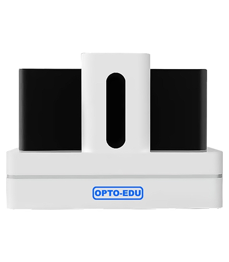
Choosing an automated microscope slide scanner is mostly a workflow decision: how many slides come in, how much detail the reader needs, and how digital slides are reviewed. OPTOEDU‘s M30 series spans compact two-slide scanning, five-slide automation, 200-slide operation, and 60/120/240/480-slide throughput.
Start with Slide Volume and Daily Workflow
Compact Scanning for Small Labs and Teaching
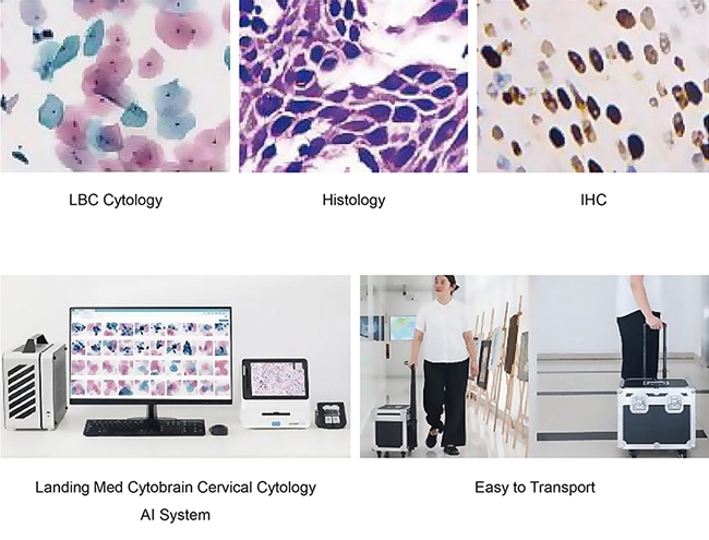
The first question is plain but often skipped: how many glass slides need to become digital slides on an ordinary day? A small laboratory, training room, or research group may only need a compact system that is easy to place, learn, and share. The M30.5832 Mini Full Auto Microscope Slide Scanner, 2 Slides is positioned for that kind of transition into digital pathology and teaching.
A compact scanner is not a compromise when the workload is modest. It can help instructors prepare teaching material, help researchers archive selected slides, and let smaller teams begin whole-slide review without building a dedicated scanning room.
Flexible Capacity for Routine Laboratory Work
Routine laboratories often sit between occasional scanning and fully automated batch scanning. The M30.1001 Digital Slide Scanner supports one to six slides and focuses on panoramic digital slide creation, browsing, area measurement, and annotation. It is a practical entry point when a team needs high-resolution digital files from traditional glass slides.
The M30.5815 Full Auto Microscope Slide Scanner, 5 Slides moves further into automated operation. It accepts five standard pathology slides or two larger pathology slides, making it suitable for smaller batches that still need automation, image stitching, and recognition functions.
High-Throughput Scanning for Large Collections
High-throughput work changes the conversation. Once a lab needs to scan trays of slides with less manual handling, capacity and unattended operation matter more than desk footprint. The M30.5840 Full Auto Microscope Slide Scanner, 200 Slides supports continuous loading and unloading, emergency slide priority, and multiple image formats such as SVS and JPG.
For larger projects, the M30.5820 Full Auto Microscope Slide Scanner, 60/120/240/480 Slides provides flexible capacity options. It fits institutions planning shared digital pathology resources or centralized teaching and research slide libraries.
Compare Automated Microscope Slide Scanner Imaging Quality
Objective Quality and Pixel Resolution
An automated microscope slide scanner has to hold enough detail for the reader to move from whole-slide context to cellular structure without fighting the image. The M30.5815 uses a plan apochromatic 20X objective with NA 0.8 and a 5.0M large-area CMOS camera. Its listed scanning resolution reaches 0.24um/pixel at 20X and 0.12um/pixel at 40X.
The M30.5820 also lists 20X and 40X scanning resolution at 0.24um/pixel and 0.12um/pixel. Pixel resolution, objective quality, illumination, autofocus, and stitching all influence digital slide review at different zoom levels.
Autofocus, Stitching, and Whole-Slide Navigation
A slide scanner does not capture a whole specimen in one simple photograph. It scans fields across the tissue area, controls focus, and stitches many images into a navigable digital slide. If stitching is poor, the reader notices it quickly: visible seams, uneven brightness, or soft detail at field edges.
OPTOEDU’s M30 products emphasize automatic scanning, high-speed scanning, tissue or contour recognition, and digital slide browsing. In M30.1001 workflows, automatic microscopic scanning and software stitching turn a traditional glass slide into a full-field digital slide. For daily users, the important outcome is smooth movement from overview to detail without losing orientation.
Use Recognition Functions to Reduce Repetitive Handling
Slide Loading and Label Recognition
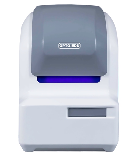
Automation earns its place when it removes the steps technicians repeat all day. The M30.5820 includes a slide loading recognition function that can skip loading points without slides during continuous scanning. It also identifies symbols, barcodes, or QR codes in slide labels.
Those functions support cleaner workflow control. A digital slide archive becomes easier to manage when label information can be connected to image records, especially in teaching, research, and consultation.
Tissue Outline and Abnormality Monitoring
Scanning the entire glass area wastes time when the useful tissue sits in one region. The M30.5820 can recognize the outline of the tissue area and skip non-organized areas during scanning. That keeps attention on the part of the slide that matters.
Machine vision monitoring for slide abnormalities adds another layer of workflow control during scanning. In a high-throughput environment, this matters because the operator cannot watch every slide manually. A system that can monitor the process gives the lab a better chance of catching positioning problems before they become unusable digital records.
Consider Browsing, File Formats, and Integration
Digital Slide Review and Annotation
The value of a digital slide is realized after scanning. Readers need to zoom, pan, annotate, measure, compare, and export useful views. The M30.1001 supports image scanning, management, and browsing software, including storage, import, annotation, marking, and teaching atlas creation.
The M30.5815 also supports real-time browsing so users can observe slide information without scanning, switch zoom ratios, capture, edit, and export images. This makes the scanner part of a review workflow rather than only a capture device.
SVS, JPG, and External Platform Compatibility
File format choices affect how easily digital slides move between users, departments, and software. The M30.5832 and M30.5840 workflows support SVS and JPG output for review, storage, and sharing needs.
Integration should still be considered case by case. A lab may need digital slide management, remote consultation software, education systems, or AI-related analysis. When remote sharing or AI platform compatibility is part of the workflow, OPTOEDU models should be matched with the actual software environment used by the laboratory.
Match OPTOEDU M30 Models to the Intended Use
M30.5832 and M30.1001 for Smaller Workloads
The M30.5832 Mini Full Auto Microscope Slide Scanner, 2 Slides suits teams that need compact brightfield scanning and a simpler path into digital slide use. The M30.1001 Digital Slide Scanner fits one to six slides, automatic focus, 20X and 40X scanning, area measurement, annotation, and full-field digital slide generation.
These systems are useful when the work is selective rather than continuous. They can support teaching sets, research slides, selected case review, and early-stage digital pathology workflows.
M30.5815 for Five-Slide Scanning
The M30.5815 is a good middle option when the lab needs automation but not a large loader. Five standard pathology slides or two large slides can be scanned with high-speed continuous area scanning. Its automatic identification functions and 20X/40X scan resolution make it suitable for smaller clinical, education, and research workflows.
M30.5840 and M30.5820 for Higher Throughput
The M30.5840 is built around 200-slide operation with continuous loading and unloading, emergency slide priority, and flexible image formats. The M30.5820 extends the discussion to 60, 120, 240, or 480 slides, matching high-volume slide libraries, institutional digital pathology projects, and large teaching collections.
Conclusion
An automated microscope slide scanner should make digital pathology easier to run, review, and scale. OPTOEDU’s M30 series gives laboratories several practical routes, from compact two-slide and six-slide scanning to five-slide automation, 200-slide operation, and flexible 60/120/240/480-slide throughput. Let the workflow lead the choice: daily slide volume, image detail, recognition needs, file handling, and review software matter before anything else.
Explore OPTOEDU’s M30 slide scanner range and contact us to build a digital slide workflow tailored to your laboratory needs.
FAQ
Q: What is an automated microscope slide scanner?
A: An automated microscope slide scanner digitizes glass microscope slides by scanning tissue areas, controlling focus, stitching image fields, and creating navigable digital slides. It reduces repeated manual microscope handling and supports review, teaching, archiving, consultation, and analysis workflows.
Q: Which OPTOEDU M30 scanner is best for a small lab?
A: A small lab may consider the M30.5832 Mini Full Auto Microscope Slide Scanner, 2 Slides, or the M30.1001 Digital Slide Scanner. The better choice depends on slide volume, software functions, scanning mode, file formats, and whether the lab wants a compact starter workflow or broader slide management.
Q: Why do slide capacity options matter so much?
A: Slide capacity affects staffing, batch planning, turnaround time, and overnight operation. A two-slide or six-slide scanner may fit selective work, while 200-slide or 60/120/240/480-slide systems better serve high-volume teaching, research, and pathology environments where unattended scanning reduces repetitive handling.
Q: What should labs check besides scan speed?
A: The following should be validated: objective quality, pixel resolution, automatic focusing, stitching quality, tissue recognition, label recognition, supported file format, browsing software, annotation capabilities, exporting options, and slide management system integration. Scan time is important, but ease of reviewing and managing the final digital slide is also important.