When a culture has to be watched for hours or days, the right live cell imaging system is the one that stays close to the cells. It should fit inside or beside the culture workflow, keep imaging conditions repeatable, capture phase contrast or fluorescence records when the assay calls for them, and make the resulting images easy to review without moving the sample back to a benchtop microscope.
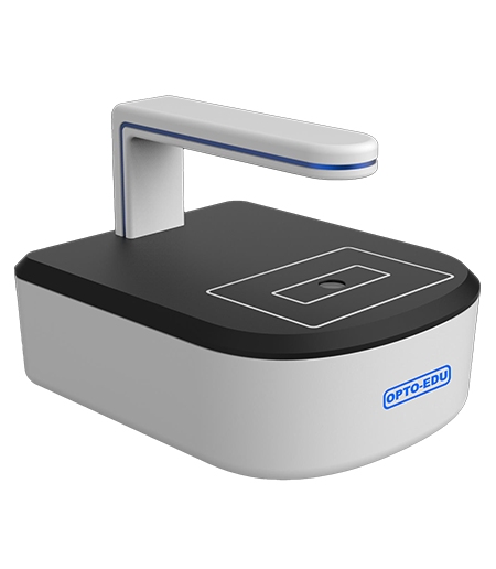
For this type of long-term observation, OPTOEDU’s M30.4810 Live Cell Imaging System provides a practical starting point. Its design supports continuous cell monitoring within the culture workflow, helping laboratories capture consistent imaging records while reducing unnecessary sample movement during extended experiments.
Why a Live Cell Imaging System Needs More Than a Camera
Protect the Culture Environment
Cell culture can be unforgiving about interruptions. Each trip out of the incubator may expose the sample to changes in temperature, humidity, gas balance, light, or contamination control. The OPTOEDU M30.4810 reduces that daily back-and-forth by supporting observation in an incubator, on a clean bench, or on a standard laboratory bench.
This matters most when the answer comes slowly. Morphology, confluence, migration, transfection, and viability may change a little at a time, then suddenly become obvious in the record. A steady imaging routine lets the researcher follow the curve instead of trying to reconstruct it from scattered snapshots.
Keep Observation Consistent Over Time
Manual microscope observation can be excellent for quick inspection, but it is harder to keep consistent across long experiments. Focus, exposure, field position, and observation timing may change from one operator to another. Automated scheduled observation gives the experiment a more repeatable rhythm.
The M30.4810 uses motorized autofocus, remote control software, and a built-in digital imaging workflow to support long-duration recording. The operator can set an observation plan and review progress remotely, which helps clean rooms and culture rooms limit unnecessary access.
Match Imaging Modes to the Biological Question
Phase Contrast for Morphology and Confluence
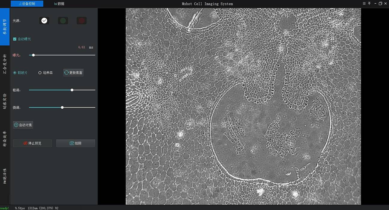
Many cell culture questions begin with morphology. Are the cells attached, spreading, dividing, rounding up, or forming a uniform monolayer? Phase contrast observation is useful because it lets researchers view unstained living cells with stronger contrast than simple brightfield observation.
The M30.4810 identifies transmitted phase contrast illumination with a 625nm LED light source and a 70mm working distance. That practical clearance helps culture vessels fit while the system still produces a usable image.
Fluorescence for Marked Cell Populations
Some experiments require fluorescence because the question is not only how cells look, but which cells are expressing a marker or responding to treatment. The M30.4810 series includes configurations with fluorescence observation, including blue and green channel options. These choices can help with transfection efficiency, cell viability workflows, and marked cell population tracking.
The important point is to choose the imaging mode around the assay. A lab focused on routine morphology and confluence may not need the same configuration as a lab tracking fluorescent labels. OPTOEDU can position the M30.4810 as a focused live-cell platform rather than a general microscope with extra accessories attached.
Turn Repeated Observation into an Analysis Workflow
Time-Lapse Imaging and Remote Alerts
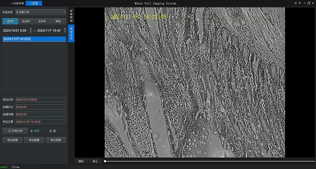
Time-lapse imaging is where a live cell imaging system becomes more than a visual tool. The M30.4810 software supports time-lapse photography, and the manual describes experiment settings such as shooting interval, shooting duration, data browsing, and time-lapse video review. The system can also support email reminders when a confluence threshold is reached.
For busy laboratories, that changes the daily routine. A researcher can follow a culture without repeatedly opening the incubator or waiting beside the workstation. The most valuable image may be the one captured at exactly the right moment, long after the first setup is complete.
Confluence, Scratch, Viability, and Transfection Analysis
For analysis, the M30.4810 software covers cell counting, cell confluence, scratch experiments, transfection efficiency, and cell viability. That shifts the work from simply collecting pictures to following measurable biological behavior. In a scratch assay, for example, the useful record is not just the open wound area on one afternoon; it is the way cells move into that space over time.
Confluence analysis is also more useful when it is measured repeatedly. A single confluence estimate may guide passaging, but a curve over time can reveal growth speed and culture condition. When images, recognition results, curves, and videos can be reviewed together, the experiment becomes easier to explain and easier to repeat.
How to Select the Right M30.4810 Configuration
Choose Between Phase Contrast and Fluorescence
Selection should begin with the sample and assay, not with the longest feature list. If the daily routine is mostly cell condition checks and growth monitoring, phase contrast may carry much of the work. Fluorescent marker tracking, viability staining, or transfection studies point toward a fluorescence-capable configuration.
The M30.4810 family includes model variants such as M30.4810-11, M30.4810-21, and M30.4810-22, with different combinations of phase contrast and fluorescence capability. The practical question is simple: which configuration answers the biological question with the least disruption to the culture?
Check Autofocus, Camera, and Working Distance
Autofocus is important because living cells may be observed across long time periods and different culture surfaces. The M30.4810 includes a motorized autofocus mechanism, and the software workflow describes coarse and fine focus adjustment as well as autofocus operation. That helps reduce the burden of manual focusing during repeated observation.
On the imaging side, the platform uses a 5.0M digital camera, while fluorescence-capable configurations can include a 2/3-inch CMOS camera and 40FPS acquisition. Frame rate, sensitivity, exposure control, and imaging mode all shape the final record. For live cell monitoring, the goal is a dependable image sequence, not one attractive still frame.
Fit the System into the Laboratory Environment
Bench Installation and Incubator Placement
A live cell imaging system has to fit into the actual room. The M30.4810 can be used indoors and is described for incubator placement, clean bench use, or standard bench use. When installed inside an incubator, the imager should be removed before a decontamination cycle, and the power supply should not be placed inside the incubator.
These details are easy to overlook during product comparison. Cable routing, workstation placement, cleaning procedure, and operator access can decide whether the instrument becomes part of the normal workflow or sits unused. A good selection process includes the sample vessel, the incubator layout, the computer, and the people who will review the data.
Remote Review and Data Management
Remote control is useful when the culture room is separate from the desk where data is reviewed. The M30.4810 software supports remote control functions including lighting control, camera control, photo capture, video capture, and data review.
Conclusion
A live cell imaging system is valuable because it keeps observation close to the culture while turning slow biological change into visible, reviewable evidence. For laboratories working with morphology, confluence, scratch assays, transfection, or viability, the OPTOEDU M30.4810 offers a practical way to reduce disturbance, automate repeated imaging, and support remote monitoring. Choose the configuration around the assay, confirm the imaging mode, and build the workflow around stable long-term observation.
Explore OPTOEDU’s live-cell imaging solution and contact us to plan a cleaner and more consistent cell monitoring process.
FAQ
Q: What is a live cell imaging system used for?
A: Labs use it to watch living cells over time while keeping the culture in a more stable environment. Typical work includes morphology checks, time-lapse imaging, confluence measurement, scratch assays, transfection tracking, and viability analysis. Its real strength is repeatable observation during ongoing cell growth.
Q: Can the M30.4810 be used inside a cell culture incubator?
A: Yes. The M30.4810 is designed for cell culture monitoring and can be placed in an incubator, on a clean bench, or on a standard bench. For incubator use, remove the imager before sterilization or decontamination cycles, and keep electrical accessories protected from moisture.
Q: When should a lab choose fluorescence live cell imaging?
A: Choose fluorescence when the experiment depends on labeled cells, transfection results, viability dyes, or specific fluorescent markers. For routine morphology and confluence, phase contrast may be enough. The deciding factor is whether the biological question needs marker-specific information.
Q: Why is motorized autofocus important in live cell monitoring?
A: Motorized autofocus helps keep images clear during repeated observation, especially when experiments run for many hours or days. It cuts down manual adjustment and supports consistent image sequences. In time-lapse work, stable focus often decides whether the final record is useful or broken.