The right laser confocal microscope should match the sample, fluorescence channels, and experimental pace. For multichannel work, the key decision is balancing wavelength coverage, detector sensitivity, optical sectioning, automation, and the need for XY, XYZ, or time-based imaging.
That balance matters in cell research, tissue observation, model-organism studies, and other workflows where one image cannot explain the sample. OPTOEDU A64 platforms offer different ways to organize the same question: how can a laboratory collect clean, repeatable fluorescence data without adding unnecessary manual steps? For laboratories comparing an automated confocal microscope with a more manual setup, repeatability is part of image quality.
Start with the Imaging Question
A laboratory should begin with the biological question rather than a list of features. Routine multicolor fluorescence on a fixed sample may need dependable excitation lines and motorized movement, while a changing sample also needs control over focus, repeated positions, exposure, and data capture.
This keeps the laser confocal microscope connected to the actual workflow. Several labels, depth information, and repeated observations call for a different configuration from a simple single-plane image, especially when the goal is 3D confocal imaging rather than a single optical section.
Match the Microscope to the Sample
Fixed Samples and Routine Multichannel Imaging
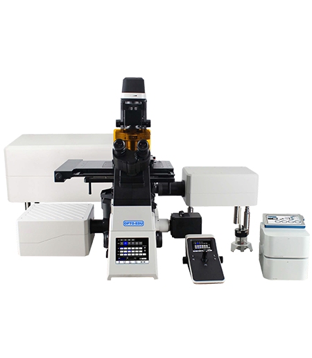
Fixed slides and prepared specimens often need dependable channel separation, clear optical sectioning, and easy movement between regions of interest. OPTOEDU A64.0960 is a practical fit when a laboratory wants a full motorized inverted fluorescence platform with 405 nm, 488 nm, 561 nm, and 640 nm laser lines. These wavelengths cover many common multichannel fluorescence arrangements without forcing the operator to rebuild the imaging path for every sample.
The A64.0960 supports XY, XYZ, and XYZT scan modes, 4K×4K scanning, 1× to 16× zoom scanning, three standard PMTs, and optional GaAsP detection. Its software supports ROI imaging, Z-stack acquisition, multi-site scanning, photobleaching, and photostimulation, helping the lab move from a broad view to a selected region and examine the same area in depth.
Live Cells and Weak Fluorescence
Live-cell experiments place extra pressure on acquisition speed and signal handling. In live cell imaging, a long observation may be affected by photobleaching, phototoxicity, focus drift, or weak fluorescence, so a suitable laser confocal microscope should help the operator shorten unnecessary illumination while keeping the image useful.
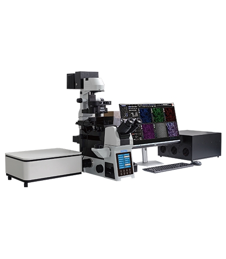
OPTOEDU A64.1020 addresses this type of workflow with high-speed resonant scanning, an array detector, four-channel PMT detection, and a 400–750 nm spectral detection range. The product page describes resonant scanning as increasing scan speed by nearly 20 times and shortening illumination time. It also describes array detection for weak fluorescence and supports X, Y, Z, λ, and T combinations for multichannel, time-lapse, multi-position, Z-stack, and panoramic imaging.
The A64.1020 also provides NIR laser confocal capability, 120 nm ultra-high resolution, motorized pinhole control, a motorized XYZ stage, and brightfield, phase contrast, polarized light, fluorescence, DIC, and Hoffman modes.
Large Samples and Repeated Acquisition
Some experiments are difficult because the sample is large, not because the individual feature is unusually small. Tissue areas, organs, and live model organisms may require a broad view, several positions, depth information, and a final image that preserves the relationship between local detail and the larger sample.
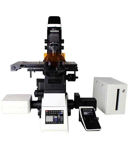
OPTOEDU A64.1010 is designed around that multi-dimensional workload. It combines four laser sources, four PMT detectors, an 8K×8K scan head, and a 25 mm confocal field of view. Its software supports multichannel fluorescence, time-lapse scanning, multi-position acquisition, Z-axis stacking, and panoramic stitching, so the imaging plan can be built around the sample rather than around one fixed field.
Evaluate the Fluorescence Workflow
Laser Channels and Spectral Flexibility
Laser lines should be selected according to the labels and the separation needed between channels. The A64.0960 offers 405, 488, 561, and 640 nm sources with AOTF wavelength control from 400 to 750 nm. A64.1010 uses four laser sources and four PMT detectors, while A64.1020 extends the workflow with five laser sources, 400–750 nm detection, four-channel PMT detection, and an array detector.
The point is not to collect every possible channel. It is to keep excitation and detection aligned with the sample so the image can distinguish the structures that matter. A clear channel plan also makes time-series and multi-position work easier to repeat.
Detector Choice for Weak Signals
Detector selection becomes more important when the signal is faint, the sample is complex, or the label cannot be used at a high concentration. Standard PMTs provide a familiar route for multichannel confocal imaging. An array detector, as used by A64.1020, is more relevant when the workflow depends on weak fluorescence and fine structural information.
The detector should be judged against signal level, optical section, required speed, and the analysis that follows. OPTOEDU gives laboratories several routes instead of making every project use the same configuration.
Z-Stack, Time Series, and Multi-Position Imaging
A useful multichannel fluorescence workflow often has more than one axis. XY imaging records a plane, XYZ adds depth, and XYZT combines depth with time. Multi-position acquisition and panoramic stitching add sample coverage when one field is not enough.
A64.0960 supports XY, XYZ, and XYZT imaging with ROI and Z-stack functions. A64.1010 and A64.1020 add combinations of X, Y, Z, wavelength, and time with multi-position, time-lapse, Z-stack, and panoramic workflows. These functions are most valuable when they reduce repeated setup and keep the same imaging logic across many locations.
Choose a Laser Confocal Microscope for Your Workflow
Choose A64.0960 when the laboratory needs a motorized inverted confocal system for routine multichannel fluorescence, flexible 3D or 4D imaging, and a familiar set of 405, 488, 561, and 640 nm laser lines. Its 4K×4K scan format and ROI tools can be a sensible balance when the sample area and data volume are moderate.
Choose A64.1010 when large sample coverage and repeated automated acquisition are central. Its 8K×8K scan head, 25 mm field of view, multi-dimensional scanning, and panoramic stitching are suited to workflows that need to connect overview imaging with detailed fluorescence analysis.
Choose A64.1020 when speed, weak-signal detection, NIR capability, and sensitive live samples carry more weight. Resonant scanning, array detection, motorized control, and the combination of fluorescence with brightfield, phase contrast, polarized light, DIC, and Hoffman observation can support a more demanding imaging routine.
Build a Repeatable Imaging Workflow
Automation is useful when it removes small variations that accumulate across a long experiment. Motorized objective switching, focusing, condenser movement, fluorescence-module changes, pinhole adjustment, and XYZ positioning can help different users follow the same sequence. The goal is not to hide the experiment behind software; it is to make the important decisions visible and repeatable.
Conclusion
Choosing a laser confocal microscope is easier when the decision follows the sample and the experiment. A64.0960 offers a flexible motorized route for routine multichannel and 3D or 4D imaging. A64.1010 is built around broad, high-detail, automated acquisition, while A64.1020 adds resonant scanning, NIR capability, and array detection for faster or weaker-signal workflows.
Explore OPTOEDU’s A64 confocal microscope range to match your fluorescence channels, sample dimensions, and imaging schedule with a platform that can grow with the laboratory’s work.
FAQ
Q: What is the most important factor when choosing a laser confocal microscope?
A: Start with the sample and the imaging question. The number of fluorescence channels, need for Z-stack or time-lapse imaging, signal strength, sample size, and required automation usually matter more than one headline resolution number. The best platform is the one that supports the complete workflow with consistent setup and useful data.
Q: Which OPTOEDU model is suitable for multichannel fluorescence imaging?
A: All three A64 models support multichannel fluorescence, but their strengths differ. A64.0960 suits routine 4K imaging with four common laser lines. A64.1010 is better for high-detail, broad-field, automated acquisition. A64.1020 is the stronger choice when weak fluorescence, NIR capability, or fast resonant scanning is important.
Q: When is an array detector useful in confocal microscopy?
A: An array detector is useful when the experiment involves weak fluorescence or fine structures that are difficult to preserve with ordinary signal levels. In the A64.1020, array detection is combined with high-speed resonant scanning and motorized control. The practical benefit depends on the sample, label, optical section, and acquisition plan.
Q: Should a laboratory choose 4K or 8K confocal scanning?
A: Choose according to sample coverage and the detail that the optical system can support. A64.0960 offers 4K×4K scanning for flexible routine work. A64.1010 provides up to 8K×8K scanning and a broad 25 mm field for larger or more detailed acquisition. More pixels are useful only when the sample and workflow can benefit from them.