In the tough field of forensic ballistics and fast-paced pathology, even the smallest mistake in imaging can cause serious problems. Lab workers and experts often face the hassle of aligning evidence by hand. Adjusting a regular stage manually tends to create shakes and focus shifts. This leads to tiredness in the body and reduces the reliability of findings. It does not matter if you aim to line up scratches on two bullet pieces or hunt for one cancer cell in a huge tissue sample. The absence of automatic accuracy remains a major holdup.
OPTOEDU knows well that your outcomes depend heavily on your tools. With more than 25 years of solid know-how and a large collection of over 1,500 microscope types, we act as a complete buying source for the globe’s pickiest labs. Our focus on strong skills makes sure that our devices, from motorized microscope stage setups to quick slide scanner devices, deliver the key features needed for exact spotting and top performance.
Anatomy of Precision: The Comparison System
For crime labs, the skill to view two items next to each other in one view area is vital. To achieve this, one needs a deep grasp of the inner structure. In addition, specific bullet comparison microscope parts make such exactness workable.
Essential Bullet Comparison Microscope Parts
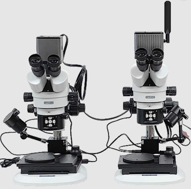
A top-quality comparison setup like the OPTOEDU A18.1830 or A18.1832 depends on various key parts to join two light paths. The core of the setup is the optical bridge. It enables left/right, split-image, and overlapping sights.
Other vital bullet comparison microscope parts cover the bullet holder set. This set lets users grip and observe all sorts of cartridges and items at varied angles and spots with ease. Moreover, a solid system must feature a magnification correction system with its own fixing lens. This part lets the operator turn a fixing dial. As a result, it ensures that two matching scales on both stages line up perfectly in overlap mode. Thus, it removes any gap between the two light paths.
The Role of the Motorized Microscope Stage in Forensic Analysis
The motorized microscope stage in the A18.1830 and A18.1832 line provides XYZ 3D motion and R turning. Users control it through a joystick or smart programs. This setup helps experts shift proof with tiny precision below a micron. Such accuracy proves crucial for matching the tiny tool lines on a bullet face. Also, a powered column offers 150mm of up/down change. Therefore, these setups can handle proof items in different sizes, from tiny threads to big cut materials.
Biological Components and Automated Imaging
In health studies, the issue moves from direct comparison to detailed scanning of large living samples. In this case, the teamwork among lenses, stages, and programs drives sharp imaging the most.
Objectives and Observation Modes
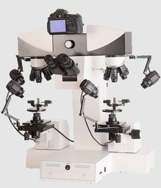
The M12 Biological and M16 Fluorescent Motorized Microscope Series suits lab and medical work. They include endless plan light setups and half-APO lenses. These setups support various viewing ways, like brightfield (BF), darkfield (DF), polarizing (PL), and fluorescence (FL). The build quality of these mechanical parts matters greatly. At the tiny scale, basic microscope changes might look slanted, which drops the focus level. OPTOEDU’s expert line guarantees a level, steady focus area over the whole sample.
High-Speed Scanning with the Slide Scanner
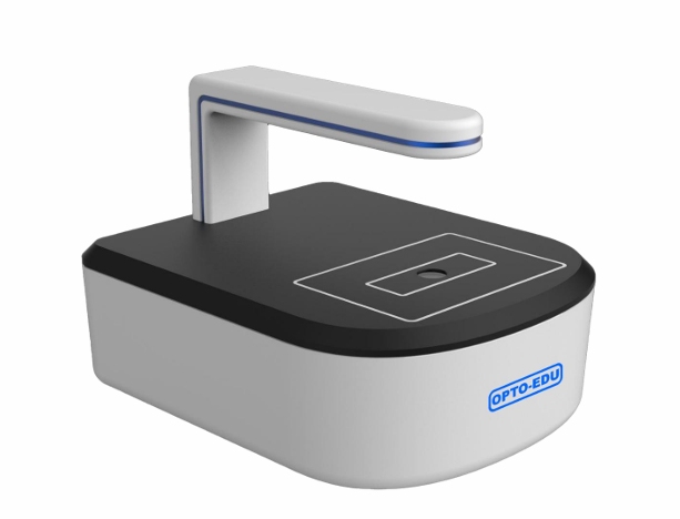
Sometimes, in order to scan and compare slices more efficiently, the slide scanner (like the M30 series) becomes essential. These devices use an auto-tiny scanning base and programs to build a complete digital slide view. A fine slide scanner manages from one slide to 480 slides in one go. It smoothly links standard glass slides into clear digital pictures.
The motorized microscope stage in these scanners pairs with clever methods. Together, they offer 2D Stitching with Auto Focus Function. This proves handy for checking samples with big height gaps. The system grabs sharp images for every field view. Then, it improves the output by fixing light shadows and line issues.
Practical Success: Case Studies
Our strong points shine brightest in the fixes we offer for tricky everyday cases.
Case Study 1: Forensic Ballistics and Questioned Documents
In crime investigation, the A18.1830 Motorized Digital Comparison Microscope helps connect bullet bits to certain guns. By using the motorized microscope stage, a specialist can spin a cartridge a full 360 degrees. At the same time, it keeps the focus steady. This lets them record tool lines exactly. The task gains from A30.1801 Professional Forensic Analysis Software. It includes tools for sizing, picture review, and image lookup. For checking doubtful papers, the setup views ink, threads, and marks next to each other. This spots fake items.
Case Study 2: Cervical Cancer Positive Cell Identification
A key issue in disease study involves scanning wide cell cuts with height gaps over 50μm. In one example, the M12.5850 motorized microscope took 5,439 images under a 40x lens. It formed a sharp, joined picture of 5.42GB. The motorized microscope stage lets the worker tap an online 2D chart for fast travel. This skips the mix-up of hard spotting typical in old high-power viewing. This fix shows how OPTOEDU tools bring exactness and speed to digital matching jobs.
Partner with OPTOEDU
Our image as a top brand rests on these solid bases. It does not matter if you seek a detailed bullet comparison microscope parts package for crime tasks or an auto slide scanner for health work. OPTOEDU gives straight factory costs and a 3-year quality promise. As the top seller for microscopes on Alibaba.com, we pledge to supply the finest picked scope for your exact needs.
Contact OPTOEDU today to boost your lab with the exactness of powered control and expert picture fixes.
FAQ
Q: What are the most important bullet comparison microscope parts for accuracy?
A: The optical bridge for side-by-side viewing, the magnification correction system, and the specialized bullet holder sets are essential for precise forensic results.
Q: How does a motorized microscope stage improve laboratory workflow?
A: It allows for sub-micron 3D movement (XYZ) and rotation, reducing manual error, preventing vibrations, and enabling automated image stitching.
Q: Can a slide scanner be used for medical diagnosis?
A: Yes, a slide scanner seamlessly stitches glass slides into digital images for full-field observation, making it ideal for pathology and research.
Q: What magnification range is available on your comparison microscopes?
A: The A18 series typically offers a wide total magnification range from 2x up to 240x, or even 288x depending on the mode.
Q: Does OPTOEDU provide software for image analysis?
A: Yes, we offer professional forensic and biological analysis software for measurement, image search, 2D/3D stitching, and image conversion.