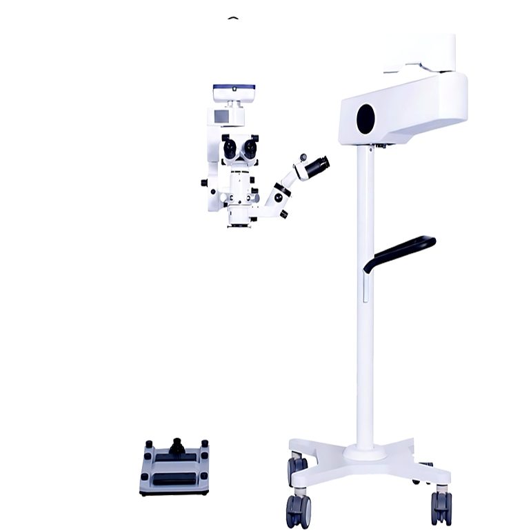
A dual-head surgical microscope is useful when a surgeon and an assistant need to follow the same ophthalmic field without relying on a compromised secondary view. OPTOEDU A41.1935 combines dual-head observation with motorized zoom, red reflection, macular protection, electromagnetic positioning, and foot-controlled operation for a coordinated eye surgery workflow.
The value of a dual-head design goes beyond adding another eyepiece. It affects teaching, supervised procedures, communication, positioning, and the way the team shares visual information. The A41.1935 dual-head ophthalmology microscope should therefore be judged by how well both observation paths support the actual work in the room.
When a Dual-Head Surgical Microscope Is Useful
Teaching and Supervised Surgery
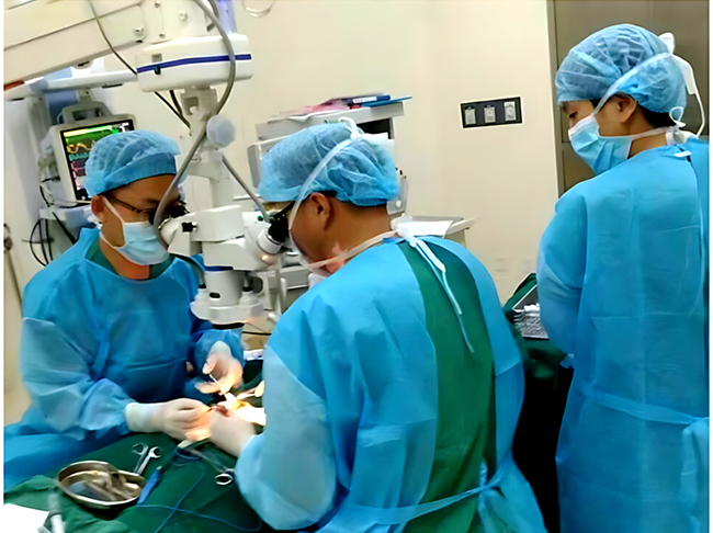
A training environment needs more than a clear image for the primary surgeon. The assistant, trainee, or supervisor must be able to follow the same field closely enough to understand the procedure and respond at the right time. A dual-head surgical microscope can support that shared view without forcing the second observer to stand away from the optical axis.
This is particularly helpful when the teaching conversation depends on small changes in the field. A matched observation path makes it easier to discuss position, instrument movement, and visual landmarks while the surgeon continues working.
Shared Observation During Team Procedures
In a collaborative procedure, assistant observation can support anticipation and communication. The second observer may need to monitor the same area, understand the surgeon’s next movement, or help explain what is visible to another member of the team. A dual-head arrangement creates a more direct visual relationship between those roles.
The purpose is not to make every procedure identical. It is to reduce the visual gap between the main surgeon and the assistant when both need a reliable understanding of the field.
How the Main and Assistant Viewing Paths Work
A Coordinated Optical Relationship
A dual-head ophthalmic surgical microscope should give the assistant a practical way to follow the primary view. A41.1935 is built around dual-head observation, red reflection, macular protection, and a continuous motorized zoom path. Together, these features help the two observers stay oriented as the field changes.
The microscope’s motorized zoom range is listed as 4.5x to 27.3x. That continuous range is useful in a shared-view workflow because the image can be adjusted gradually rather than shifting abruptly between fixed magnification steps.
Adjusting Viewing Angles and Position
Different surgeons and assistants do not share the same height, posture, or preferred viewing angle. A usable dual-head system must allow the optical arrangement to be positioned without creating a strained working posture. A41.1935 combines an adjustable dual-head design with a balanced cantilever structure and a mobile stand.
The practical test is simple: can the main observer and assistant both maintain a clear, comfortable view while the microscope remains close to the surgical field? If the answer depends on repeated manual repositioning, the dual-head feature will not deliver its full value.
Positioning and Ergonomics in the Operating Room
Balanced Movement with Electromagnetic Locks
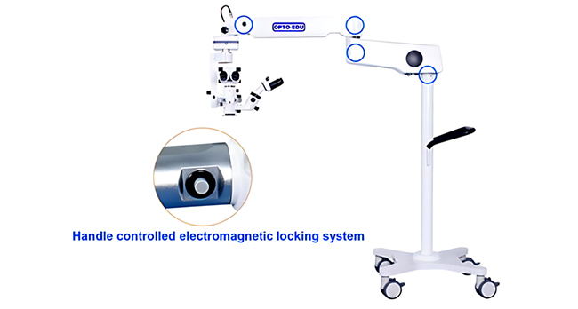
A41.1935 uses three electromagnetic locks to support free-floating, balanced positioning. The handle button releases the lock system so the microscope lens can be moved toward the working area. Releasing the button locks the joints again, helping the microscope remain stable once the team has established the view.
For a surgical microscope for eye surgery, this movement pattern matters because the optical head must be positioned accurately without making the surgeon fight the stand. Quick release and dependable locking can make it easier to adjust the microscope between procedures or when the surgical field changes.
Adapting the System to Different Users
In teaching hospitals and shared operating rooms, the microscope may be used by clinicians with different working habits. Foot-controlled zoom, focus, X-Y movement, brightness, and image functions reduce the need to reach toward the optical head. That helps each user make small changes while keeping the hands in the working zone.
The assistant observation microscope concept is therefore tied to ergonomics. The second viewing path has to remain useful when the primary surgeon changes posture, magnification, focus, or position.
Extending Observation with a BIOM Kit
Non-Contact Wide-Angle Fundus Viewing
A41.1935 can be paired with an optional BIOM kit for fundus and vitreoretinal surgery. The system provides non-contact wide-angle viewing with a panoramic, high-resolution view of the retina without corneal contact.
For a BIOM surgical microscope, the main benefit is a broader posterior-segment view that can be shared within the ophthalmic workflow. This gives specialist rooms another way to think about the dual-head platform, especially when the team needs a common view of the vitreous, retina, or macular area.
A Broader Workflow for Specialist Rooms
The BIOM option does not mean every facility needs the same configuration. It means the microscope can be evaluated in relation to the procedures the room actually supports. If the workflow includes posterior-segment observation, accessory compatibility becomes as important as the basic dual-head structure.
Features to Check Before Selecting a Dual-Head Microscope
Optical Alignment and Magnification
Start by checking whether the main and assistant observers can maintain a useful relationship with the field as magnification changes. The core dual-head surgical microscope decision should include zoom range, red reflection, illumination, focus, and the ability to keep the field readable for both observers.
Foot Control and Image Capture
The A41.1935 foot switch controls motorized zoom, focus, X-Y movement, brightness, image capture, recording, browsing, freeze, light control, page movement, and filter switching. Those functions can support a teaching workflow in which the team needs to pause, capture, review, or explain a view without repeatedly touching the optical head.
Stand Movement and Working Space
The microscope should fit the room’s access paths, table position, and operator posture. A mobile stand can help with placement, but the relevant question is whether the microscope can move into the correct position and then remain stable. The assistant’s physical location and the clearance around the stand should be considered during setup.
OPTOEDU A41.1935 for Collaborative Ophthalmic Workflows
OPTOEDU A41.1935 brings dual-head viewing together with motorized continuous zoom, dual coaxial LED illumination, red reflection, macular protection, electromagnetic locks, and a multifunction foot switch. That combination suits rooms where observation is shared between a surgeon and assistant, or where teaching and documentation are part of the daily workflow.
The product is most relevant when the facility needs a stable common view rather than a primary microscope plus a disconnected secondary observation method.
Conclusion
A dual-head surgical microscope earns its place when shared observation improves the way a team teaches, communicates, positions the microscope, and follows an ophthalmic procedure. OPTOEDU A41.1935 supports that role through dual-head viewing, continuous 4.5x-27.3x motorized zoom, electromagnetic locks, hands-free foot control, red reflection, macular protection, and an optional BIOM kit for fundus and vitreoretinal surgery.
Contact OPTOEDU to discuss how the A41.1935 configuration can match your operating room’s optical and positioning requirements.
FAQ
Q: What is the main benefit of a dual-head surgical microscope?
A: The main benefit is shared observation. A surgeon and assistant, trainee, or supervisor can follow the same ophthalmic field more closely than they could with a separate, poorly aligned viewing method. This supports communication, teaching, coordinated movement, and a clearer understanding of the procedure.
Q: Is a dual-head microscope useful for cataract surgery training?
A: It can be useful when a trainee or supervisor needs to follow the primary surgeon’s view during instruction. The value depends on the optical alignment, viewing comfort, magnification control, and the way the room conducts teaching. A41.1935 combines dual-head observation with motorized zoom and foot control.
Q: What does the optional BIOM kit add?
A: The optional BIOM kit adds non-contact wide-angle viewing for fundus and vitreoretinal surgery. It is described as providing a panoramic, high-resolution retinal view without corneal contact. The accessory is most relevant to facilities whose ophthalmic workflow includes posterior-segment observation.
Q: Which features should be checked before choosing a dual-head microscope?
A: Check the optical relationship between the two viewing paths, zoom and focus control, illumination, red reflection, positioning locks, foot switch functions, stand movement, image capture, and accessory compatibility. The best choice is the one that keeps both observers comfortable and the surgical field stable.