Confocal microscopy resolution is not determined by pixel count alone. A useful evaluation must connect scan sampling with the objective, numerical aperture, pinhole, detector, field of view, and the size or movement of the sample. That is why a 4K confocal system can be the better choice for one workflow, while an 8K system is more useful for another.
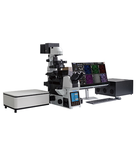
The OPTOEDU A64 range makes this trade-off visible. A64.0960 provides 4Kx4K scanning across a 19 mm field of view, while A64.1010 and A64.1020 support up to 8192×8192 scanning across a 25 mm confocal field. The difference is not simply a higher number; it changes how a laboratory balances coverage, detail, acquisition time, and data volume.
How Confocal Microscopy Resolution Should Be Evaluated
Separate Pixel Sampling from Optical Resolution
Scan size describes how many pixels are used to represent the selected field. Moving from 4K to 8K increases digital sampling across that field, which can preserve more image information when the optical system and sample contain enough detail. It does not automatically double the microscope’s optical resolving power.
Optical resolution is also shaped by wavelength, objective numerical aperture, pinhole size, alignment, signal quality, and the specimen itself. A larger file can still look soft if the objective, focus, or signal-to-noise ratio limits the image before it reaches the detector.
Consider the Objective and Detection Path Together
A confocal system rejects much of the light outside the focal plane, which improves section contrast and supports 3D reconstruction. The result depends on the complete optical path. High-NA objectives can collect more fine detail, while a suitable detector and controlled pinhole help preserve useful signal after optical sectioning.
For this reason, confocal microscopy resolution should be evaluated as a workflow outcome. Ask whether the final image can distinguish the structure, layer, boundary, or fluorescence pattern that matters to the experiment, rather than treating scan pixels as a stand-alone quality score.
Why Field of View Changes the Imaging Strategy
The 19 mm Field of View of A64.0960
A64.0960 offers a 19 mm confocal field of view with 4Kx4K scanning and 1x to 16x zoom scanning. That combination gives the operator room to frame the sample broadly, then increase scan zoom for a selected region. It is useful when a laboratory moves between whole-area context and more focused 3D or fluorescence observations.
The 25 mm Field of View of A64.1010 and A64.1020
A64.1010 and A64.1020 use a 25 mm confocal field of view and support up to 8192×8192 scanning. The broader field can reduce the need to divide a large sample into as many separate views, while the larger scan matrix gives more pixels to describe that area.
The value becomes clearer with tissue sections, large cell areas, or multi-position experiments. A broad field can preserve context, while motorized stages, Z-stacks, and panoramic stitching help the user connect local detail with the larger specimen.
When 4K Is Enough and When 8K Helps
A64.0960 for Flexible 4K Scanning
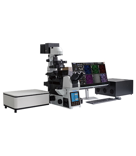
A 4K system can be the right answer when the field, objective, and sample detail are already well matched. OPTOEDU A64.0960 combines 4096×4096 scanning with four laser lines at 405, 488, 561, and 640 nm, three standard MA PMTs, and an optional GaAsP PMT. Its software supports XY, XYZ, XYZT, ROI, and multi-site imaging.
For a laboratory that needs routine multicolor fluorescence, 3D cell culture observation, histological sections, or flexible region-of-interest work, 4K can provide a sensible balance between image detail, file size, and acquisition time.
A64.1010 for High-Detail Automated Imaging
A64.1010 is a stronger fit when up to 8192×8192 scanning and automated acquisition are central requirements. Its 25 mm field of view supports broad sample coverage, while multidimensional functions such as Z-stack, multi-position, time-series, and panoramic workflows help organize repeated imaging.
The advantage of 8K is most practical when the optical system can support the added sampling and the sample contains useful structure across the selected field. Otherwise, the larger file may increase storage and review demands without adding meaningful information.
A64.1020 for 8K Speed and Weak Signals
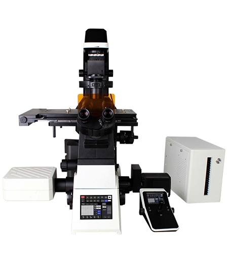
OPTOEDU A64.1020 combines up to 8192×8192 scanning with resonant scanning, array detection, and an optional 730 nm NIR laser path. The platform is aimed at workflows where high-detail sampling must coexist with rapid acquisition or faint fluorescence. Its 25 mm field of view also supports broader observation before the operator moves into a selected region.
The model is therefore not simply an 8K alternative. It is a choice for laboratories that need to manage scan speed, signal recovery, spectral information, and sample coverage together.
Balance Resolution with Sample Dynamics
Thick Samples Need Section Control
Thick samples often contain signals above and below the focal plane. Confocal optical sectioning reduces the contribution of that out-of-focus light, while XYZ scanning turns separate optical planes into a depth-resolved record. The purpose is not to create a large image file; it is to preserve spatial relationships through the sample.
Dynamic Samples Need Time Awareness
Time-lapse imaging adds a second dimension to the resolution decision. A very large scan may capture detail but take too long for a changing sample. Resonant scanning on A64.1020 can support faster acquisition, while the other A64 platforms provide multidimensional modes that let users choose a more controlled balance.
Match Detail to the Experimental Question
The best setting is the lowest scan and optical burden that answers the question clearly. A large tissue region may need a wide field and stitching. A small subcellular structure may need a higher-NA objective, a tighter optical section, and more pixels across a smaller area. A repeatable workflow keeps these decisions visible rather than defaulting to the largest available number.
A Practical Selection Rule for Research Labs
Choose by Sample Coverage
Use the 19 mm A64.0960 field when the sample and workflow are comfortably covered by that area or when the lab expects to work through ROI and zoom scanning. Consider the 25 mm fields of A64.1010 and A64.1020 when broader context, large sections, or fewer stitched positions are important.
Choose by Scan Dimension
If the experiment depends on depth, compare XYZ and XYZT capability with the actual acquisition plan. If it depends on multiple labels, include wavelength handling and detector channels. If it depends on repeated locations, check motorized stage and multi-position control rather than judging resolution from pixel count alone.
Choose by Detection and Future Expansion
Detector choice becomes important when fluorescence is weak or spectrally close. A64.1020 adds array detection and optional NIR capability, A64.0960 offers MA PMT and optional GaAsP detection, and A64.1010 provides an automated 8K platform for high-detail imaging. The right choice is the one that leaves room for the samples the lab actually expects to study.
Conclusion
Good confocal microscopy resolution comes from balancing sampling, optical sectioning, detector performance, field of view, and acquisition time. OPTOEDU A64.0960 offers flexible 4K imaging across a 19 mm field, while A64.1010 and A64.1020 extend the scan matrix and field to 8K and 25 mm, with A64.1020 adding resonant scanning and array detection.
Review the OPTOEDU A64 confocal microscope range and match the platform to the sample coverage and imaging dimensions that matter most.
FAQ
Q: Does 8K automatically mean twice the optical resolution of 4K?
A: No. 8K provides more sampling pixels across the selected field, but optical resolution also depends on wavelength, objective numerical aperture, pinhole, alignment, detector performance, and sample quality. The added sampling is most useful when the optical path and specimen contain detail that can support it.
Q: When is a 4K confocal microscope a practical choice?
A: 4K can be practical when the sample area, objective, and imaging question are well matched to the available sampling. OPTOEDU A64.0960 combines 4Kx4K scanning with four laser lines, motorized control, ROI imaging, XYZ and XYZT modes, and 1x to 16x zoom scanning for flexible research workflows.
Q: Why does field of view matter in confocal imaging?
A: Field of view determines how much of the sample can be framed in one acquisition. A wider field can preserve context and reduce the number of stitched positions, while a smaller field may be easier to use for focused ROI imaging. A64.0960 uses a 19 mm field, while A64.1010 and A64.1020 use 25 mm fields.
Q: Which A64 model is suited to thick or changing samples?
A: Thick samples benefit from optical sectioning and XYZ imaging, while changing samples also require attention to acquisition time and exposure. A64.1020 is the strongest fit when resonant scanning and weak-signal detection are central. A64.0960 and A64.1010 remain useful when flexible multidimensional control is the main need.