In the challenging environment of biological science research, there is no room for mistakes. Scientists frequently encounter situations where they have to work with inadequate imaging tools that create blurred images, high levels of background noise, and phototoxicity. Such issues go beyond creating bad images since they result in misinterpretation of information and delayed scientific advancements. For high-stakes laboratory environments, the need for a laser confocal microscope for laboratory research that offers uncompromising precision is paramount.
OPTOEDU bridges this gap between research requirements and technological capability. OPTOEDU has been operating for more than 25 years in the international market of microscopes and has positioned itself as a leading supplier of top-of-the-line optical systems. Our main aim is to develop high-performing systems that allow researchers to investigate the nanoscale world without any difficulty.
The Fundamental Principle of the Confocal Microscope
Understanding what the principle of the confocal microscope is is essential for appreciating why it has become the gold standard for biological imaging. Unlike traditional widefield microscopy, which captures light from the entire specimen, confocal systems utilize a spatial pinhole to eliminate out-of-focus light.
Optical Conjugation and Signal Integrity
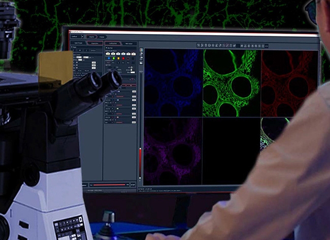
The core principle of confocal microscope technology lies in the conjugate relationship between the light source, the sample, and the detector. In the OPTOEDU A64.1010 system, the pinhole design is based on the principle of light reversibility. Both the excitation beam and the emitted light from the specimen will travel through the same computer-controlled pinhole, thus making sure that they remain completely conjugated to each other at 100%. The novel approach helps filter out the non-focal plane light much more efficiently.
Achieving Super-Resolution Imaging
By utilizing high-precision confocal microscope optics for rapid sample imaging, our systems can achieve scanning resolutions up to 8192×8192 pixels. This level of detail allows scientists to capture vital biological data with unprecedented efficiency, transforming how we visualize complex tissues and organ systems.
Advanced Detection: Precise Confocal Microscopy Detection for Living Cells
Live-cell imaging presents a unique set of challenges, primarily the need to balance high-speed acquisition with the preservation of cell viability. Traditional systems often subject sensitive samples to prolonged laser exposure, leading to photobleaching and cell death.
How Confocal Microscopy Detection Works for Live Cell Imaging
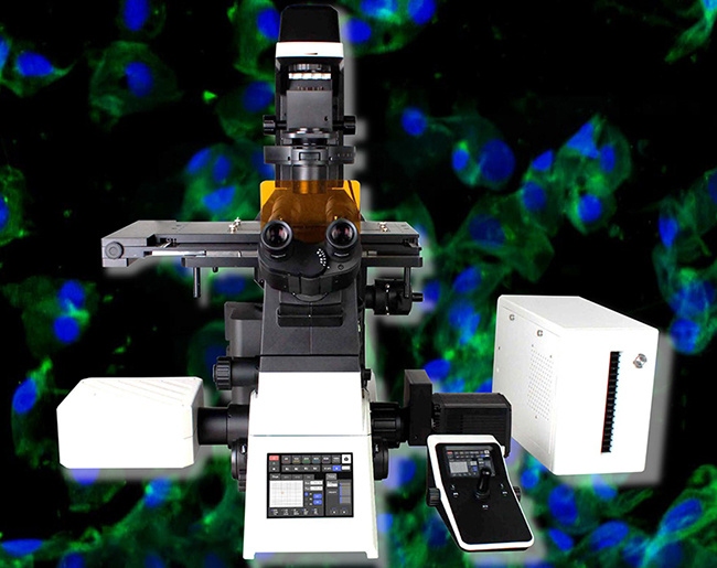
To address this, the OPTOEDU A64.1020 incorporates resonant scanning technology. But how confocal microscopy detection works for live cell imaging in our high-end models involves more than just speed; it involves the intelligent management of light. Resonant scanning increases frequencies by nearly 20 times, drastically shortening illumination time while maintaining exceptional image quality. This process reduces phototoxicity, facilitating the precise confocal microscopy detection for living cells over extended periods without compromising their natural state.
Low-Noise Array Detection
Our laser confocal microscope for laboratory research utilizes space array detection technology. By employing several closely packed SPAD detector elements, the system captures two-dimensional spatial data at each scanning point. This enables the acquisition of ultra-fine features that are otherwise not captured by conventional confocal detection methods, thus making it possible to obtain very clear images from extremely faint fluorescent signals.
High-Throughput Applications inbiological science Research
Modern biological research demands a deeper and broader analysis of model organisms. Whether it is oncology, neuroscience, or developmental biology, the ability to extract more data from a single sample is a competitive advantage.
Expanding Field of Vision
The A64.1010 model boasts an industry-leading 25mm field of view. This allows for the capture of large samples in a single scan, offering 1.5 times the data throughput compared to standard systems. When combined with high-precision confocal microscope optics for rapid sample imaging, researchers can analyze entire tissues or organs with high efficiency, reducing the need for time-consuming manual stitching.
Multi-Dimensional Imaging Modes
Life science experiments often require complex observation parameters. Our systems support the combined use of X, Y, Z, λ, and T scanning functions. Researchers can freely combine multi-channel fluorescence imaging, time-lapse scanning, and Z-axis stacking to adapt to diverse experimental scenarios.
Case Analysis: High-Precision Imaging in Modern Oncology
In a recent study involving tumor microenvironments, researchers utilized the OPTOEDU A64.1020 to track the migration of cancer cells in real-time. By leveraging precise confocal microscopy detection for living cells, the team was able to observe delicate cellular interactions over a 48-hour period.
The system’s resonant scanning minimized photobleaching, allowing for continuous high-resolution imaging of weak fluorescent markers. Moreover, the NomisProX-C software was used to control the hardware effectively, enabling the scientists to concentrate on the biological implications of the experiment and not worry about complicated microscope procedures. This is an example of how an advanced laser confocal microscope in the laboratory can help speed up the process of medical discoveries.
Why Choose OPTOEDU as Your Strategic Partner?
For brand owners and distributors looking for where to buy a laser confocal microscope with high-resolution imaging, OPTOEDU offers more than just hardware. We provide a comprehensive ecosystem of support, including a 3-year quality warranty and direct factory expertise.
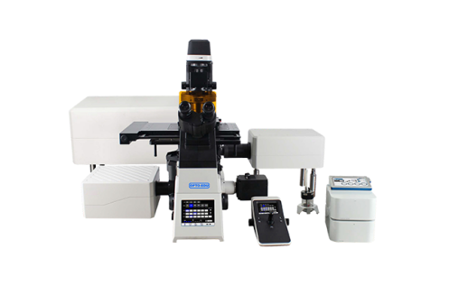
Our hardware is designed for “Super Vision, Super Resolution, and Super Speed”. From motorized nosepieces and condensers to intuitive touch-screen interfaces, every element of our laser confocal microscope for laboratory research is engineered to be an extension of the researcher’s own eyes and hands.
Conclusion
The impact of confocal microscopy onbiological science research is profound, but the quality of the instrument determines the validity of the result. By choosing OPTOEDU, you are investing in high-precision confocal microscope optics for rapid sample imaging and a legacy of optical excellence. Our commitment to high-precision detection ensures that your laboratory is equipped to handle the most challenging frontiers of biological science.
Contact OPTOEDU today to explore our full range of confocal solutions and elevate your research capabilities.
Email: sale@optoedu.com
Mobile / Wechat: +86 13911110627
WhatsApp: +86 1088696020
Address: F-1501 Wanda Plaza, No.18 Shijingshan Road, Beijing 100037, China
FAQ
Q: Why is the field of view 25 mm in your confocal system useful?
A: It allows double the amount of information gathering, since scientists can scan larger samples in one go.
Q: How does the A64.1020 system prevent any damage to living cells?
A: Resonant scanning helps increase speed up to 20 times, thus reducing the exposure time of the laser beam and its effect on cells.
Q: Where can I purchase a laser confocal microscope with high-quality imaging and expert assistance?
A: OPTOEDU provides top-notch confocal microscopes worldwide with 25+ years of experience and a 3-year warranty.
Q: Is it possible to configure scanning modes according to experimental requirements?
A: Yes, we offer multi-dimensional scanning (X, Y, Z, λ, T), which can be freely configured for advanced studies.