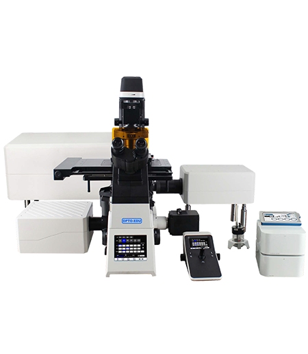
The pursuit of clarity of microscopic samples led to the development of high-tech imaging systems that go far beyond the conventional widefield microscope. Confocal microscope is the top-of-the-line tool in modern scientific research and offers a high-contrast image of three-dimensional specimens. By means of precise lasers and sensitive detectors, the samples can be explored non-destructively in great detail, especially for complex biological material as well as for man-made materials.
Defining the Precision of the Laser Confocal Microscope
The main advantage of laser confocal microscope is that it can be used to exclude out-of-focus light. This is often called optical sectioning and is in contrast to wide-field microscope, where a whole sample is illuminated, and the light from above and below the in-focus part is collected. The laser confocal microscope uses a point source of light and a conjugate pinhole in front of the detector to collect only light from the in-focus part of the sample.
The Role of Spatial Pinholes in Optical Sectioning
The spatial pinhole is the critical component that defines the “confocal” nature of the system. By matching the aperture of the pinhole to the size of the Airy disk formed by the objective lens, the system rejects signal from outside the focal volume. This precision results in images with significantly higher signal-to-noise ratios, allowing for the observation of fine details that would otherwise be obscured by background haze.
Elevating Biological Research Through Fluorescence Imaging
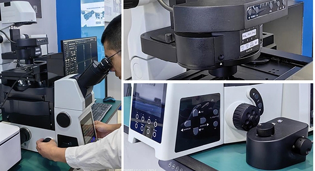
When confocal microscope is mentioned in life sciences, high-end fluorescence imaging is not far. This technology is extremely valuable for localizing molecules within cells and tissues by determining their spatial distribution. For scanning confocal microscopes, this is achieved by moving a laser beam focused in the specimen plane as a raster to map the fluorescent signals with extreme spatial accuracy and to create a high-resolution image.
Visualizing Intracellular Dynamics and Localization
Fluorescent probes can be attached to proteins, nucleic acids, and organelles. Because the scanning confocal microscope can be set to a thin optical section, samples with multiple labels can be observed clearly without the colors from the different depths ‘bleeding’ into each other. This is especially critical for studies of co-localization of proteins within microscopic compartments where it is necessary to establish whether two proteins interact, i.e., are found within the same compartment.
- Mapping the distribution of cytoskeletal elements.
- Tracking the movement of vesicles during endocytosis.
- Study of the integration of viral genetic material into the host nucleus.
Achieving Depth with 3D Structure Analysis
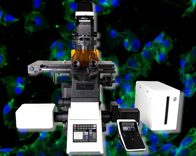
The capability to perform 3D reconstructions of the specimens is one of the most important features of confocal microscope. For this purpose, the specimen is scanned point by point through the focal plane, a process known as Z-stacks. These 2D images are then reassembled by software into a 3D representation that can be viewed from all sides, measured, and even analyzed with respect to certain parameters.
From Z-Stacks to Volumetric Reconstruction
By viewing volumetric reconstructions of objects that have been imaged in 3D, one can gain insight and information that cannot be gleaned from viewing 2D representations of an object. In neurobiology, for example, the 3D structure of neurons and their complex branching enables a precise connection map of neurons. In materials science, for example, laser confocal microscopes are used to investigate internal cavities and grain boundaries of translucent samples. All of the internal structure of the sample can be viewed without physically sectioning the sample.
Technological Frontiers of the OPTOEDU A64 Series
The OPTOEDU A64 series is the cutting-edge technology in integrated imaging, for both research in universities and quality control in industry. The systems run automatically, completely, and continuously, without human intervention. Full-auto for full complexity of imaging.
A64.1020: Pioneering NIR and Full Auto Capabilities
OPTOEDU A64.1020 is a flagship NIR (Near-Infrared) laser confocal microscope system. It extends the capability of traditional imaging methods. Because Near-Infrared light penetrates deeply into samples, it enables us to image thick samples, such as brain sections or organoids. OPTOEDU A64.1020 is equipped with high-performance Apochromatic (APO) objectives, which assure the best color correction and image flatness over the entire field of view.
A64.1020 is designed for laboratory use and supports all necessary observation modes.
- Brightfield (BF) and Phase Contrast (PH) for standard morphology.
- Polarizing (PL) and Fluorescence (FL) for molecular and structural information.
- Differential Interference Contrast (DIC) and Hoffman Modulation Contrast to view transparent specimens.
A64.1010 and A64.0960: Streamlining Motorized Workflows
OPTOEDU’s flagship for NIR imaging is joined by two more models for fluorescence and DIC imaging: A64.1010 APO full-auto and the A64.0960 scanning confocal for specialized motorized applications: BF, PL, FL, and DIC imaging with XYZ and nosepiece motorized scanning over large areas to collect data fast from big samples.
Expanding Horizons in Materials Science and Industrial Inspection
The Laser Confocal Microscope has many prominent biological applications, but it is also a highly valuable tool for the semiconductor and materials industries. By performing non-contact 3D surface metrology, the confocal laser can measure the surface of micro-components with vertical resolution in the nanometer range. OPTOEDU supports this field with our A64 series as well as with special systems such as the M20 motorized industry microscopes.
Surface Metrology and Non-Destructive Testing
OPTOEDU’s M20.0910 and M20.3820 models are perfect for 3D measurement and super-depth-of-field (EDF) analysis. The surface roughness of components, wear on mechanical parts, as well as the integrity of micro-fabricated circuits can be characterized with these industrial systems. They work with the principle of confocal microscope and therefore provide much more detail than a usual optical inspection system. With these systems, the highest quality is measured and checked.
Conclusion
Integration of confocal microscope into today’s research laboratory has brought profound change. From the requirements of high-resolution fluorescence imaging in the molecular biological laboratory through to the requirement for precise 3D structure analysis in the engineering materials laboratory, the scanning confocal microscope remains the method of choice for cutting-edge research. OPTOEDU A64 series of products provides a sophisticated automated high-precision instrument suite designed to enable users to advance the frontiers of their fields of study.
Contact us to learn how we can support your advanced imaging requirements.
FAQ
Q: How does the pinhole in confocal microscope improve image quality?
A: In confocal microscope, the pinhole acts as a spatial filter. It is placed in the conjugate focal plane of the microscope, allowing light from the focal plane to pass through while blocking out-of-focus light from other depths. This ensures that the scanning confocal microscope captures only the sharpest part of the image, significantly increasing clarity and contrast compared to widefield systems.
Q: What are the main benefits of the OPTOEDU A64.1020 NIR system?
A: The OPTOEDU A64.1020 NIR laser confocal microscope has the unique ability to image near-infrared, allowing for increased depth of penetration into specimens and reduced chance of phototoxicity. Paired with the full-auto capabilities of the system and the APO objectives, the NIR laser confocal microscope provides high-quality, deep-tissue 3D structure analysis, particularly suited to thick tissues and live organoids.
Q: What imaging modes are available in the OPTOEDU A64 series?
A: The OPTOEDU A64 series is a versatile multi-modal microscope. The different models, like the A64.1020, can be used for Brightfield (BF), Phase Contrast (PH), Polarizing (PL), Fluorescence (FL), Differential Interference Contrast (DIC), and Hoffman Modulation Contrast on one laser confocal microscope system.
Q: Is the OPTOEDU A64.0960 suitable for automated high-throughput imaging?
A: The OPTOEDU A64.0960 is a full-auto motorized scanning confocal microscope that has been designed for maximum efficiency in the laboratory. The OPTOEDU A64.0960 is motorized for fully automated control of the XYZ axes as well as the nosepiece. As such, it is an ideal tool for any high-throughput automated workflow. Even working manually, it is designed to enable the user to take identical high-quality images of many samples and even of very large areas of individual samples with a minimum of manual repositioning.