In the demanding area of cell biology, clear visualization often sets the line between a key discovery and an unsuccessful test. Numerous labs and business allies face issues with basic widefield microscopy. There, stray light from unfocused areas causes a constant haze that hides vital details inside cells. Such poor sharpness results in wrong results, lost materials, and delayed advances. To overcome these vision limits, the sector needs exact tools that can explore the intricacies of live organisms with remarkable detail.
As a top confocal microscope supplier in China, OPTOEDU builds the link from fuzzy views to tiny-scale sharpness. With more than 25 years of solid know-how, we have become a main ally for premium optical tools. Our focus stays on helping scientists via better spotting accuracy and smooth automated steps.
Understanding Precision: The Foundation of Confocal Technology
To grasp how cutting-edge setups change biological studies, you should first tackle the basic query: what is a confocal microscope? In essence, the confocal microscope definition describes an optical method that boosts image sharpness and depth by employing a tiny pinhole to cut off unfocused light during sample viewing.
The Laser Confocal Microscope Principle
The key strength of this approach rests on the laser confocal microscope principle. Traditional scopes light up the whole specimen at once, but a confocal one uses laser beams spot by spot. Its setup relies on the idea of light reversal: both the exciting beam and the glow from the sample go through the same adjustable pinhole, which keeps a full matching link.
This setup makes sure that just the light from the main focus point gets picked up. As a result, it blocks unwanted glow from other levels. For a confocal microscope supplier in China, such as OPTOEDU, fine-tuning this pinhole with smooth, powered adjustments proves crucial. It helps reach top spotting power and picture clarity.
Applications in Modern Cell Biology
Knowing what a confocal microscope is used for matters a lot for business partners aiming to equip tough labs. These devices go beyond simple viewers; they serve as versatile study hubs.
Deep Tissue and Live-Cell Imaging
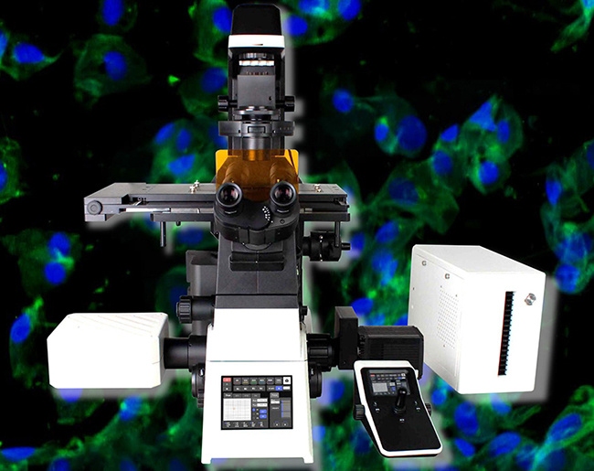
Current studies call for closer looks at tissues, body parts, and test creatures. Tools like the OPTOEDU A64.1010 support varied imaging across axes (X, Y, Z, λ, T). They enable layer-by-layer views in Z and wide joins for big samples. This proves especially useful for:
- Time-lapse scanning:Watching active cell changes as time passes.
- Thick specimen analysis:Getting sharp pictures from deep inside living forms, where normal light would spread out.
Reducing Phototoxicity with High-Speed Scanning
A major hurdle in watching live cells involves phototoxicity, which is harm to cells from long laser contact. Our main model, the A64.1020 NIR Laser Confocal Microscope, tackles this via quick resonant scan methods. These boost scan rates by about 20 times. Thus, fast capture keeps picture standards high while cutting exposure time a lot. This setup aids extended views of delicate live cells, all without risking their health.
The OPTOEDU Advantage: Technical Highlights and Detection Accuracy
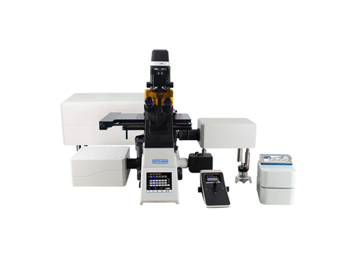
As a skilled confocal microscope supplier in China, OPTOEDU has crafted its A64 line to fit the strictest needs for fast and fine imaging. We stress spotting exactness and part blending so our trade partners get top-tier gear.
Superior Field of View and Resolution
The A64.1010 offers a leading 25mm view area, which gives 1.5 times more data flow than older types. Paired with a scan sharpness up to 8192×8192 pixels, it lets experts pull richer details from one pass with amazing fineness.
Advanced Detection Technology
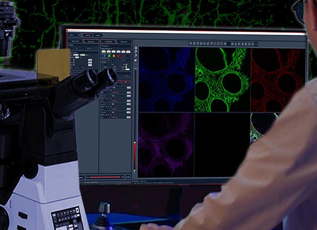
Spotting exactness marks the core of the OPTOEDU name. The A64.1020 employs area-based spotting tech with several nearby SPAD sensors. This group gathers flat spatial data at every scan spot. Consequently, it brings back super-small features reaching 120nm sharpness that usual confocal spotting often misses. This fine spotting makes sure even faint glow signals come through clearly.
Seamless Hardware and Software Integration
Lab speed comes from auto features. Our setups include total powered handling of the lens holder, XYZ platform, light adjuster, and glow wheel. The NomisProX-C program runs this, linking hardware commands and picture review tightly. As an “all-in-one” choice, it spares scientists from heavy-handed work, so they can center on their core ideas.
Why Partner with OPTOEDU?
For company leaders and sellers, picking a confocal microscope supplier in China means more than checking specs; it involves trust and name value. OPTOEDU ranks high on global sites like Alibaba.com. We earn praise for steady service and strong quality checks, including a 3-year cover on our lab-level tools.
We deliver premium, study-ready options that match worldwide tops, providing:
- Extreme Precision:Quick resonant scans and sensor arrays.
- Full Automation:Screen-based controls and powered parts for easy flows.
- Comprehensive Support:Skill in picking ideal scope setups for given study goals.
By adding OPTOEDU’s sharp confocal tech to your range, you give clients the means to push biology limits and escape the haze of old imaging ways.
Prepared to lift your offerings with exact confocal tools? Reach out to OPTOEDU now to talk about teaming chances and tech details.
FAQ
Q: What is a confocal microscope?
A: It is a special optical setup that uses a space pinhole to remove unfocused light, giving better sharpness and depth than regular scopes.
Q: What are the main benefits of the laser confocal microscope principle?
A: The principle enables “optical slicing,” which lets you grab clear pictures from set depths in thick samples by holding a matching tie via a pinhole.
Q: Why choose OPTOEDU as your confocal microscope supplier in China?
A: We bring 25+ years of skill, exact items like the A64 line with 120nm sharpness, full auto features, and a 3-year quality promise.
Q: What is a confocal microscope used for in live-cell research?
A: It serves for quick, varied imaging of live cells, causing minimal light harm while grabbing active biology steps in real time.