AFM surface roughness measurement becomes more efficient when the operator can see the target before scanning it. An optical microscope provides the visible context and two-dimensional measurement route, while AFM supplies three-dimensional height information from the selected area. An integrated platform keeps those steps close together, which is the central value of an optical atomic force microscope.
This matters for thin films, coatings, particles, microstructures, and samples that need to be observed in liquid. The goal is not to replace every other microscope; it is to connect an optical view of the sample with a local AFM measurement that explains how the surface is shaped.
How an Optical Atomic Force Microscope Supports AFM Surface Roughness Measurement
From Visible Target to AFM Measurement Area
Surface analysis often starts with a feature that is visible at a larger scale: a coating edge, a particle, a scratch, a patterned region, or a change in texture. The challenge is to move from that visible target to a smaller AFM scan without losing the location or relying on repeated trial and error.
An optical atomic force microscope addresses that transition by placing optical navigation and probe-based scanning in one working relationship. The operator can locate the region through the optical path, then use AFM to examine local height, texture, and roughness.
Why 2D and 3D Data Answer Different Questions
Optical 2D measurement is useful for dimensions, edges, visible patterns, and the broader position of a feature. AFM 3D measurement adds height information, so the result can show whether a region is raised, recessed, stepped, or rough in a way that a flat optical image cannot fully describe.
For a coating study, the optical image may show coverage and a visible defect, while the AFM scan can help quantify the local surface profile. The two data types are strongest when they describe the same area and support the same decision.
What A62.4505 Adds to an Integrated Imaging Workflow
Simultaneous Optical and AFM Imaging
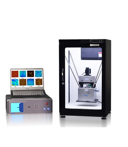
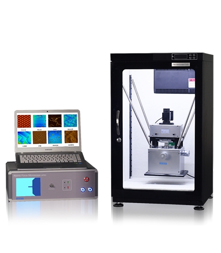
The OPTOEDU A62.4505 optical atomic force microscope combines an optical metallographic microscope and an atomic force microscope in one integrated design. Its optical and AFM imaging functions can work at the same time without affecting each other, allowing the operator to maintain visible context while collecting local surface information.
That continuity is useful when the feature is small, the sample has several similar regions, or the surface must be revisited after the first scan. The optical view helps keep the measurement grounded in the physical sample instead of leaving the operator with an isolated nanoscale image.
Optical 2D Measurement and AFM 3D Measurement
A62.4505 supports optical 2D measurement and AFM 3D measurement in the same platform. For a laboratory, this creates a practical division of labor: use the optical channel for orientation and visible dimensions, then use the AFM channel when height, roughness, or local morphology becomes the important question.
The value is especially clear when the lab needs to compare what a feature looks like with what its surface profile actually is. Instead of treating the optical and AFM results as unrelated files, the operator can build a more coherent interpretation of one sample region.
When Liquid Sample AFM Analysis Matters
Using the Sample Liquid Cell
Some surfaces are most meaningful in a liquid environment. Drying can change the way a specimen is arranged or how a soft surface behaves, so a laboratory may need to observe the sample while it remains covered by the test liquid.
A62.4505 includes a liquid sample cell workflow in which liquid is introduced through the liquid tank of the probe holder until it covers the probe and glass. This makes the platform relevant to liquid sample AFM analysis when the sample, probe, and measurement method are compatible with that setup.
Observation During Liquid Injection
An integrated optical view can make that preparation easier to follow because the operator is not working entirely without visual context. The final method still depends on the sample and the selected cell arrangement, but the combined platform supports a more direct observation-to-measurement sequence.
Where an All-in-One AFM Can Improve Laboratory Work
Thin Films, Coatings, and Surface Texture
Thin films and coatings often need two kinds of evidence. Optical observation can show coverage, edges, patterns, and larger defects, while AFM can reveal roughness, local steps, particles, and fine texture. For an AFM surface roughness measurement workflow, the integrated platform is useful when the same region needs both visual context and a three-dimensional profile.
Microstructures and Local Surface Features
Microstructures can be easy to find optically but difficult to interpret without height information. A visible ridge, depression, or patterned feature becomes more informative when the AFM result shows its local topography.
The same logic applies to particles and surface contamination. Optical imaging helps identify where a feature sits, while AFM can help distinguish a raised particle from a color or contrast change. This makes the integrated workflow suitable for targeted investigation rather than broad, unfocused scanning.
How A62.4505 Relates to Routine and Research-Level Options
When A62.4501 Is Enough for Routine Measurement
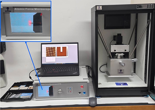
Not every surface measurement needs an optical-AFM combination. The OPTOEDU A62.4501 basic AFM platform is a reasonable fit when the laboratory already knows the scan area and mainly needs contact or tapping measurements within a compact range. Its listed 20 um scan range and nanoscale resolution values support routine work when the sample fits the measurement window.
When A62.4503 Needs Research-Level Flexibility
The OPTOEDU A62.4503 research AFM system is more relevant when the surface question changes from project to project. Selectable piezoelectric scanners, precision probe positioning, automatic approach, optical positioning, and environmental monitoring support a research workflow that may need to explore different regions and conditions.
A62.4505 is the stronger choice when the central challenge is preserving the connection between optical context and AFM measurement. A62.4503 is the stronger direction when research flexibility and controlled probe-based exploration matter more than integrated optical 2D measurement.
Selection Questions for an Optical AFM Workflow
Ask whether the operator needs to find a visible target, measure it optically, and then scan the same area in three dimensions. If the answer is yes, an integrated AFM imaging system can reduce the gap between the first observation and the final surface result.
Why OPTOEDU Makes Integrated AFM Workflows More Practical
AFM surface roughness measurement is most useful when the measured area still has a clear meaning. OPTOEDU A62.4505 connects optical observation, optical 2D measurement, AFM 3D measurement, and a liquid-cell workflow in one platform. That combination can help laboratories move from “What is this feature?” to “What is its actual surface profile?” with fewer disconnected steps.
The result is a more grounded approach to surface analysis: locate visually, measure locally, and interpret the two views together. Explore the OPTOEDU A62.4505 integrated AFM solution when your laboratory needs a clearer path between optical inspection and nanoscale surface data.
Conclusion
An AFM surface roughness measurement workflow becomes stronger when optical context and AFM topography answer the same surface question. OPTOEDU A62.4505 brings optical 2D measurement, AFM 3D measurement, simultaneous imaging, and liquid sample support into one integrated platform, while A62.4501 and A62.4503 remain useful when the priority is compact routine work or research-level flexibility.
Contact us to discuss which OPTOEDU AFM configuration best fits your surface measurement and research requirements.
FAQ
Q: What is the main advantage of an optical atomic force microscope?
A: An optical atomic force microscope connects visible sample observation with AFM-based surface measurement. The optical channel helps locate and understand a target, while AFM adds local three-dimensional height and texture information. This is useful when the same region needs both optical context and nanoscale topographic analysis.
Q: How do optical 2D measurement and AFM 3D measurement differ?
A: Optical 2D measurement is suited to visible dimensions, edges, patterns, and location. AFM 3D measurement records surface height and local morphology, helping describe roughness, steps, particles, ridges, and depressions. Together, the two methods explain both where a feature is and how its surface is shaped.
Q: Can A62.4505 support liquid sample AFM analysis?
A: A62.4505 includes a liquid sample cell workflow in which liquid is introduced through the probe holder until it covers the probe and glass. This supports liquid-state observation when the sample, probe, and method are compatible with the cell arrangement. The final procedure should be matched to the specimen and experiment.
Q: When is A62.4501 a better choice than A62.4505?
A: A62.4501 can be the better choice when the laboratory has a known scan region and needs routine contact or tapping measurements in a compact range. A62.4505 is more valuable when optical location, optical 2D measurement, AFM 3D measurement, or liquid-cell observation must work together in one platform.